Overview of Rectoanal Polyps in Dogs
Rectoanal polyps are flap-like growths arising from the mucosal lining of the rectum or anus in dogs. These growths (also called rectal polyps) are typically benign adenomatous lesions – essentially extensions of the intestinal lining – that can either attach by a narrow stalk or have a broad base. Most rectoanal polyps remain non-cancerous; however, they carry a risk of malignant transformation over time. Larger polyps have a greater chance of harboring or developing into malignant tumors (adenocarcinoma).
This article is written by VOSD Advance PetCare for the reference of both veterinarians and pet parents. If this is an emergency, you should refer to your nearest vet. This article is a recommendation of the treatment for the condition in reference and is based on drugs and medication available in India. Doses & administration should strictly be administered by a trained professional vet. VOSD is in no way liable for the use of this information for the dog(s) in your care. For more information, you can reach the VOSD Advance PetCare services, including VOSD AI or the VOSD CloudVet, both available via the VOSD website Please note that these services are free for dog owners and subscription-based for vets and vet hospitals.
No single definitive cause has been identified for rectal polyps. They are generally considered idiopathic (of unknown origin), though middle-aged to older dogs are more commonly affected. There is no strong breed or sex predilection noted. Chronic inflammation in the colorectal region is suspected as a possible contributing factor in some cases, but evidence is inconclusive. At VOSD Advance PetCare, our veterinary team often encounters these polyps in senior dogs during routine exams or when investigating signs of straining and bleeding. Each case is approached with the understanding that while benign polyps are manageable, vigilance is needed due to the potential for recurrence or malignancy.
Symptoms and Clinical Signs
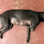
Dogs with rectoanal polyps may present with a variety of lower intestinal signs, often related to the physical presence of the polyp in the rectum. Key clinical signs include:
Tenesmus (Straining to defecate)
Affected dogs commonly strain or exhibit pain while passing stool. They may assume the posture to defecate frequently with little result.
Hematochezia
Fresh red blood in the feces is a hallmark sign. The blood is often seen on the surface of otherwise normal or slightly firm stool, due to the polyp’s fragile surface bleeding.
Mucus in Stool
The feces may be coated in mucus. Polyps irritate the rectal lining, leading to excess mucus production.
Ribbon-like or Thin Stools
If a polyp is large enough to partially obstruct the rectal lumen, fecal matter can appear pencil-thin or flattened. This occurs when the mass narrows the passage for stool.
Painful Defecation (Dyschezia)
Dogs might vocalize or show discomfort when defecating. Owners may mistake this for constipation or hemorrhoids, but in dogs, it often indicates a rectal lesion.
Visible Mass
In some cases, a soft, red protruding mass may be seen at the anus, especially during or after defecation. Polyps are typically well-vascularized, friable, and may ulcerate, so any protruding tissue can look inflamed and bleed on touch. Occasionally, the polyp prolapses through the anal orifice during defecation and then retracts.
Not all signs are present in every case – for example, a small polyp may only cause mild intermittent bleeding with minimal straining. However, any dog with recurrent hematochezia, straining, or mucus in stool should raise suspicion for a rectal mass among other differentials.
Diagnosis of Rectoanal Polyps in Dogs
Diagnosing rectoanal polyps requires a combination of clinical examination and specific diagnostic procedures performed by a veterinarian. Because the symptoms overlap with other anorectal conditions, a thorough approach is essential for accurate diagnosis. Key steps in the diagnostic workup include:
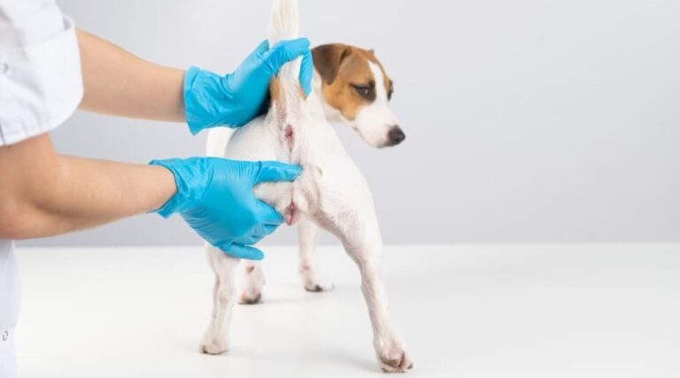
Physical and Rectal Examination:
A complete physical exam is performed first, paying special attention to a digital rectal examination. Often, the polyp can be directly palpated as a mass in the distal rectum, and it tends to bleed easily upon touch. If the polyp is near the anal opening, it may even be visible on exam. This exam also helps assess the size, texture, and number of masses present. For a comfortable and thorough rectal exam, sedation may be used especially if the dog is in pain.
Colonoscopy (Endoscopic Evaluation)
Colonoscopic examination is the gold standard for visualizing rectoanal polyps. Using a flexible endoscope, the veterinarian can inspect the entire rectum and colon for polyps or other abnormalities. This allows identification of single vs. multiple polyps and assessment of their exact location and attachment.
Notably, routine abdominal X-rays or ultrasound are usually not very useful to detect polyps directly (small soft-tissue polyps won’t show up on radiographs and may be missed on ultrasound). However, imaging can be employed to rule out other issues or check regional lymph nodes if a malignant tumor is suspected.
Biopsy and Histopathology
Definitive diagnosis hinges on histopathological examination of the polyp tissue. During colonoscopy, the veterinarian will biopsy the polyp or, more often, remove it entirely (polypectomy) for lab analysis. Histopathology confirms whether the lesion is a benign adenomatous polyp or a malignant growth (such as carcinoma). A biopsy sample should always be submitted for diagnosis, since benign polyps and early malignancies can appear similar externally. The histology will also reveal if there are any precancerous changes at the margins.
Laboratory Tests
Routine lab work, while not diagnostic for polyps specifically, is important for a complete evaluation. A Complete Blood Count (CBC) and biochemistry profile are typically normal in isolated rectal polyps, but they are useful to assess the dog’s overall health (e.g. check for anemia from chronic bleeding, or organ function prior to anesthesia). In addition, a fecal examination is often done to rule out parasitic or infectious causes of bleeding that could mimic polyp symptoms.
If chronic bleeding or ulceration is present, testing coagulation profiles might be considered to ensure normal blood clotting before surgery.
Differential Diagnosis
It is crucial to distinguish rectoanal polyps from other conditions that produce similar signs. Anal gland abscesses, rectal strictures, inflammatory masses, or rectal prolapse can also cause tenesmus and bleeding. More importantly, malignant tumors must be ruled out – these include carcinoma in situ or rectal adenocarcinoma, and other neoplasms like leiomyomas, lymphomas, or papillomas in the rectal area.
Polypoid adenomas are benign, but an early carcinoma can appear polypoid, so only histology can differentiate them. If a malignant tumor is suspected (for example, a very firm or invasive-feeling mass), additional diagnostics like chest radiographs or abdominal ultrasound should be done to check for metastasis, as part of the staging process.
By systematically performing these diagnostic steps, a veterinarian can confirm the presence of rectoanal polyps and assess their nature. In practice at VOSD Advance PetCare, we ensure that every rectal mass is thoroughly evaluated endoscopically and histologically, so that an optimal treatment plan can be formulated with no surprises. Early and accurate diagnosis allows for prompt intervention before polyps cause serious complications.
Treatment and Management of Rectoanal Polyps in Dogs
Surgical removal is the cornerstone of treatment for rectoanal polyps in dogs. In fact, definitive cure is achieved by excising the polyp tissue, which not only resolves the current symptoms but also prevents the benign growth from turning malignant over time. Medical management alone is generally insufficient to eliminate a polyp, though it can aid in symptom relief and post-operative care. Below is a step-by-step outline of the management of rectoanal polyps, including specific procedures and supportive therapies (with commonly available medications in India):
Preoperative Preparation
Proper preparation ensures a safer surgery and recovery. The dog should be stabilized and evaluated before anesthesia. This includes completing any needed diagnostic tests (as above), correcting dehydration or other underlying issues, and fasting the dog prior to the procedure. Many veterinarians also perform a bowel preparation – for example, administering a stool softener or an enema the day before surgery – to clear the rectum of feces. A clean distal colon reduces the risk of infection during surgery by minimizing fecal contamination. Additionally, a broad-spectrum antibiotic is often started prior to surgery as prophylaxis (especially because the rectal surgery site is not sterile). In India, a common choice would be an antibiotic like amoxicillin-clavulanate (e.g. Clavamox® or Augmentin®) started the day of surgery to prevent post-operative infection.
Surgical Polyp Removal (Polypectomy)
The primary treatment is to excise the polyp completely. The approach depends on the polyp’s size and location:
For polyps near the anal opening or in the distal rectum, a trans-anal surgical excision can often be performed. Under general anesthesia, the veterinarian gently everts the anus (or uses a speculum) to expose the polyp and then excises it with surgical instruments, obtaining as much of the stalk or base as possible. The mucosal defect may be sutured closed if large, or left to heal by second intention if small.
For polyps located further inside the rectum or if multiple polyps are present, an endoscopic polypectomy is preferred. Using a colonoscope, the vet can deploy a wire snare to lasso and cut the polyp at its base (often with electrocautery). This minimally invasive technique achieves removal without an open surgical incision, resulting in less trauma and quicker recovery. At our facility, we favor endoscopic removal whenever feasible, as it allows excellent visualization and access to polyps that are out of reach on manual exam.
In cases of a very broad-based (sessile) polyp or suspected malignancy, a more extensive surgery might be required. This can involve a partial proctectomy or full-thickness resection of the affected rectal segment, sometimes approached via abdomen or by a rectal pull-through procedure. Such invasive surgery is rare for benign polyps, but if the polyp is confirmed as malignant (adenocarcinoma) on biopsy, wider surgical margins are necessary to achieve cure. Referral to a surgical specialist for these advanced procedures is recommended when cancer is involved.
During polyp removal, care is taken to minimize bleeding. The rectal tissue is well-vascularized, so precise technique and cautery are used to achieve hemostasis. If the polyp is removed endoscopically, an electrocautery snare often helps in cauterizing blood vessels as the stalk is cut. After excision, the surgical site is examined for any remnant tissue. Intraoperative colonoscopy may be repeated to ensure no additional lesions are missed higher up in the colon. Each excised polyp is immediately placed in formalin and sent for histopathology.
Biopsy and Histopathologic Confirmation
Every polyp that is removed must be submitted to a pathology laboratory for microscopic examination. This step is critical – it distinguishes benign polyps from malignant tumors definitively. The histopathology report will identify if the lesion is an adenomatous polyp (benign) or if there are areas of carcinoma within it. It will also comment on whether surgical margins appear clean (in a pedunculated polyp, the margin is at the stalk base). Benign rectal adenomas are usually cured by complete excision. However, if the report indicates a malignancy or atypical cells at the margins, further surgical intervention or adjunct therapy (such as a wider resection or even chemotherapy) may be indicated. Thus, the treatment plan can be adjusted once the biopsy results return, underlining why confirming the diagnosis via histology is so important for proper cure.
Treatment and Management of Rectoanal Polyps in Dogs
Surgical removal is the cornerstone of treatment for rectoanal polyps in dogs. In fact, definitive cure is achieved by excising the polyp tissue, which not only resolves the current symptoms but also prevents the benign growth from turning malignant over time. Medical management alone is generally insufficient to eliminate a polyp, though it can aid in symptom relief and post-operative care. Below is a step-by-step outline of the management of rectoanal polyps, including specific procedures and supportive therapies (with commonly available medications in India):
Preoperative Preparation
Proper preparation ensures a safer surgery and recovery. The dog should be stabilized and evaluated before anesthesia. This includes completing any needed diagnostic tests (as above), correcting dehydration or other underlying issues, and fasting the dog prior to the procedure. Many veterinarians also perform a bowel preparation – for example, administering a stool softener or an enema the day before surgery – to clear the rectum of feces. A clean distal colon reduces the risk of infection during surgery by minimizing fecal contamination. Additionally, a broad-spectrum antibiotic is often started prior to surgery as prophylaxis (especially because the rectal surgery site is not sterile). In India, a common choice would be an antibiotic like amoxicillin-clavulanate (e.g. Clavamox® or Augmentin®) started the day of surgery to prevent post-operative infection.
Also read: Chronic Inflammation of the Anus, Rectum or Perineum Region in Dogs
Surgical Polyp Removal (Polypectomy)
The primary treatment is to excise the polyp completely. The approach depends on the polyp’s size and location:
For polyps near the anal opening or in the distal rectum, a trans-anal surgical excision can often be performed. Under general anesthesia, the veterinarian gently everts the anus (or uses a speculum) to expose the polyp and then excises it with surgical instruments, obtaining as much of the stalk or base as possible. The mucosal defect may be sutured closed if large, or left to heal by second intention if small.
For polyps located further inside the rectum or if multiple polyps are present, an endoscopic polypectomy is preferred. Using a colonoscope, the vet can deploy a wire snare to lasso and cut the polyp at its base (often with electrocautery). This minimally invasive technique achieves removal without an open surgical incision, resulting in less trauma and quicker recovery. At our facility, we favor endoscopic removal whenever feasible, as it allows excellent visualization and access to polyps that are out of reach on manual exam.
In cases of a very broad-based (sessile) polyp or suspected malignancy, a more extensive surgery might be required. This can involve a partial proctectomy or full-thickness resection of the affected rectal segment, sometimes approached via abdomen or by a rectal pull-through procedure. Such invasive surgery is rare for benign polyps, but if the polyp is confirmed as malignant (adenocarcinoma) on biopsy, wider surgical margins are necessary to achieve cure. Referral to a surgical specialist for these advanced procedures is recommended when cancer is involved.
During polyp removal, care is taken to minimize bleeding. The rectal tissue is well-vascularized, so precise technique and cautery are used to achieve hemostasis. If the polyp is removed endoscopically, an electrocautery snare often helps in cauterizing blood vessels as the stalk is cut. After excision, the surgical site is examined for any remnant tissue. Intraoperative colonoscopy may be repeated to ensure no additional lesions are missed higher up in the colon. Each excised polyp is immediately placed in formalin and sent for histopathology.
Biopsy and Histopathologic Confirmation
Every polyp that is removed must be submitted to a pathology laboratory for microscopic examination. This step is critical – it distinguishes benign polyps from malignant tumors definitively. The histopathology report will identify if the lesion is an adenomatous polyp (benign) or if there are areas of carcinoma within it. It will also comment on whether surgical margins appear clean (in a pedunculated polyp, the margin is at the stalk base). Benign rectal adenomas are usually cured by complete excision. However, if the report indicates a malignancy or atypical cells at the margins, further surgical intervention or adjunct therapy (such as a wider resection or even chemotherapy) may be indicated. Thus, the treatment plan can be adjusted once the biopsy results return, underlining why confirming the diagnosis via histology is so important for proper cure.
Postoperative Care and Medications of Rectoanal Polyps in Dogs
After removal of the polyp, appropriate post-op care ensures the dog heals comfortably and complications are avoided. Key aspects of management in the recovery period include:
Pain Management
Rectal surgery can be mildly painful, so analgesics are administered. Non-steroidal anti-inflammatory drugs (NSAIDs) are commonly used to control pain and inflammation. In India, veterinarians may prescribe carprofen (brand name Carodyl®, Rimadyl®, etc.) or meloxicam (Melonex® oral suspension) for a few days post-surgery. These NSAIDs help the dog feel more comfortable during defecation. For very sensitive patients, additional pain relief (e.g. tramadol) can be given as needed.
Antibiotics
Even if started before surgery, antibiotics are usually continued for several days afterwards to guard against infection. The rectum is a non-sterile field, and despite cleaning, bacteria can invade the surgical site. A broad-spectrum antibiotic effective against colonic flora is chosen. Examples include metronidazole (effective against anaerobes in the colon) often combined with a fluoroquinolone, or a broad agent like amoxicillin-clavulanate. The exact choice depends on the surgeon’s preference and the dog’s health status.
Stool Softeners and Diet
To protect the healing rectal mucosa, it’s vital to keep the dog’s stools soft and easy to pass. Stool softeners such as lactulose syrup are routinely prescribed. Lactulose (e.g. Duphalac® in India) is a gentle laxative that draws water into the bowel, keeping feces soft. It can be given orally (typically 1 ml/2 kg dose, adjusted to effect) starting immediately after surgery.
Alternatively or additionally, docusate sodium (a stool softening agent) can be used. A high-fiber, bland diet is recommended for a week or two – this could include a prescription intestinal diet or homemade low-residue food – to ensure smooth bowel movements. The goal is to prevent any straining or constipation that could tear sutures or cause bleeding.
Local Care
In most cases, the surgical site is internal and not visible externally, so local wound care is minimal. If any part of the polyp was external or if the anus was incised, the vet may advise keeping the area clean with a dilute antiseptic rinse. An Elizabethan collar (E-collar) is used to stop the dog from licking or traumatizing the anal region as it heals.
The first bowel movement after surgery is often delayed a day or two due to pre-surgery fasting and enemas. When it occurs, some minor blood spotting is normal, but the stool should be soft. Owners are instructed to observe the dog closely and report any excessive bleeding, severe pain, or straining.
Must Read: Breast Cancer in Dogs (Mammary Gland Tumours
Follow-up and Prognosis
Postoperative follow-up is extremely important in cases of rectoanal polyps. The treating veterinarian should re-examine the surgical site about 10–14 days after surgery to ensure proper healing. This visit often includes a gentle rectal exam to check for swelling or infection (and to remove any sutures if non-absorbable sutures were placed at the anus). Subsequently, periodic rechecks are recommended because polyps can recur.
A common schedule is to perform rectal exams at 3 months and 6 months post-surgery, and thereafter every 6–12 months. Regular surveillance is aimed at catching any new polyps early, since new polyps may develop after surgery in some dogs. Dogs that had a single, benign polyp and clean margins tend to have an excellent prognosis – many never experience a recurrence. In contrast, dogs with multiple polyps or diffuse polyposis have a higher risk of recurrence. If new polyps are found on follow-up, they can be removed endoscopically before they grow large.
In the case of polyps that were found to be malignant, additional therapy and vigilant monitoring (including imaging for metastasis) will be part of the ongoing management, and the prognosis will depend on the extent of disease.
Overall, the prognosis for dogs with rectoanal polyps is very good when appropriate treatment is given. Surgical or endoscopic polyp removal is typically curative for benign polyps, resulting in rapid relief of symptoms and a return to normal comfort during defecation. Dogs usually recover quickly – often showing improvement in stool quality and comfort by the time of their two-week check-up. Long-term survival is excellent for benign cases; many dogs live out their normal lifespan without significant issues related to the condition. The key is diligent follow-up to catch any new growth early.
Conclusion
In summary, rectoanal polyps in dogs are a condition that, while relatively uncommon, should be on the radar for any veterinarian evaluating an older dog with rectal bleeding or straining. With a careful diagnostic approach and timely surgical intervention, these polyps can be removed before they cause serious harm. As this guide outlines, modern veterinary practice – including at VOSD Advance PetCare – emphasizes minimally invasive removal techniques, appropriate adjunct medications, and thorough follow-up care to ensure the best outcome. By treating rectal polyps proactively, we can vastly improve a dog’s quality of life and prevent benign conditions from progressing to something more dangerous.