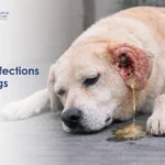
Testicular cancer in dogs is one of the most common cancers in older, unneutered male dogs and one of the most treatable when caught early. In rescue work, it is frequently encountered in dogs who have never been neutered, sometimes living with tumours that have been present and growing for months without detection. The encouraging reality is that most dogs treated with timely surgery make a full recovery and go on to live comfortable lives. Understanding what testicular cancer looks like, what causes it, and what can be done about it is the first step towards protecting your dog.
What Is Testicular Cancer in Dogs?
Testicular cancer in dogs occurs when abnormal cells develop and multiply within one or both testicles. The testicles have multiple cell types, each of which can give rise to a different kind of tumour. This is why a dog can develop more than one type of testicular tumour simultaneously a finding that is actually quite common in clinical practice.
Unlike many internal cancers, testicular tumours in dogs are often detectable through simple physical examination, which means an observant pet parent or a routine veterinary check-up can identify the problem before it becomes advanced.
Types of Testicular Tumours in Dogs
There are three main types of testicular tumours diagnosed in dogs:
- Sertoli cell tumour: Arises from the cells that support and nourish sperm development. Sertoli cell tumours are particularly significant because they can produce oestrogen, causing feminising changes in the dog such as hair loss, enlarged mammary glands, and attraction from other male dogs. They are more commonly found in retained (undescended) testicles.
- Seminoma: Arises from the sperm-producing cells of the testicle. Seminomas are generally considered to have low malignant potential and rarely spread to other organs, though exceptions do occur. They tend to be the most common type in normally descended testicles.
- Leydig cell tumour (interstitial cell tumour): Arises from the cells that produce testosterone. These tumours are typically small, slow-growing, and benign. They rarely cause clinical problems beyond the local enlargement they create.
All three types can occur together in the same dog. Only laboratory analysis of the surgically removed tissue can confirm the exact type and guide any follow-up care needed.
Symptoms of Testicular Cancer in Dogs
Because the testicles are accessible externally, testicular cancer in dogs is often identifiable during routine grooming or bathing if pet parents know what to look for. Common signs include:
- Swelling or enlargement of one or both testicles
- A noticeable asymmetry between the two testicles one significantly larger than the other
- A firm, irregular, or lumpy texture to the affected testicle
- Hair loss on the flanks, belly, or around the groin area (often associated with hormone-producing Sertoli cell tumours)
- Enlargement of the mammary glands (nipples becoming more prominent)
- Attraction from other intact male dogs due to elevated oestrogen levels
- A pendulous or unusually shaped prepuce (foreskin)
- Lethargy and reduced activity levels
- Difficulty walking or sitting comfortably if the testicle has become very large
- In dogs with a retained testicle, abdominal swelling or discomfort may be the first noticeable sign, as the undescended testicle is hidden inside the body
Make a habit of gently feeling your unneutered male dog’s testicles during bath time or grooming. Any change in size, shape, or texture should prompt a veterinary visit without delay.
Causes and Risk Factors
Testicular cancer in dogs is associated with several well-established risk factors:
- Age: The condition is most commonly diagnosed in dogs above 9 to 10 years of age. The risk increases progressively as dogs get older.
- Cryptorchidism (retained testicle): This is the single most significant risk factor for testicular cancer in dogs. When one or both testicles fail to descend into the scrotum and remain inside the abdominal cavity or inguinal canal, the risk of developing a tumour particularly a Sertoli cell tumour or seminoma is dramatically higher than in normally descended testicles. Cryptorchid dogs should be neutered as early as possible to eliminate this risk.
- Lack of neutering: Intact male dogs carry a lifelong risk of testicular cancer that neutered dogs do not. Early castration removes this risk entirely.
- Breed predisposition: Afghan Hounds, Boxers, German Shepherds, Shetland Sheepdogs, and Weimaraners are among the breeds reported to have higher incidence rates, though testicular cancer can occur in any breed.
Finding testicular cancer in your dog is not caused by anything you did or did not do. However, it is worth knowing that timely neutering is the most effective preventive measure available.
Related Videos


How Vets Diagnose Testicular Cancer
Diagnosis of testicular cancer in dogs is usually straightforward when the testicles are in the scrotum. The process typically involves:
1. Physical Examination: The vet will carefully palpate both testicles, assessing size, symmetry, texture, and tenderness. Asymmetry or a firm, irregular mass is strongly suggestive of a tumour.
2. Ultrasound: Scrotal ultrasound provides a detailed view of the internal structure of the testicle and can help identify the extent and nature of the mass. For dogs with retained testicles, abdominal ultrasound is used to locate the undescended testicle and assess it for tumour development.
3. Blood Tests: A full blood count and biochemistry panel assess overall health and evaluate for changes associated with hormone-producing tumours including anaemia caused by oestrogen excess from Sertoli cell tumours.
4. Imaging for Spread: Chest X-rays and abdominal ultrasound are performed to check whether the cancer has spread to the lymph nodes or lungs, which is uncommon but important to rule out before surgery.
5. Biopsy and Histopathology: The definitive diagnosis is made after surgical removal of the affected testicle(s). The tissue is sent to a laboratory where the tumour type is confirmed and any features suggesting aggressive behaviour are identified.
Treatment Options for Testicular Cancer
The treatment for testicular cancer in dogs is, in most cases, straightforward and highly effective:
1. Castration (Surgical Removal of the Testicles): Surgical removal of both testicles is the primary and most effective treatment. This eliminates the tumour, removes the source of any abnormal hormone production, and prevents new tumours from developing in the remaining testicle. In dogs with a retained testicle, the surgical approach will include exploration of the abdomen or inguinal region to locate and remove it.
For the majority of dogs with testicular cancer that has not spread, castration alone is curative. Most dogs recover quickly from the procedure and return to normal activity within one to two weeks.
2. Chemotherapy: Rarely required for testicular cancer in dogs. It may be considered in the uncommon situation where the cancer has already spread to lymph nodes or other organs at the time of diagnosis.
3. Supportive Treatment for Hormonal Effects: Dogs with oestrogen-producing Sertoli cell tumours may have developed bone marrow suppression as a result of prolonged oestrogen exposure a condition that can cause life-threatening anaemia. These dogs may require supportive care including blood transfusions before or after surgery. Hormonal changes typically reverse after the tumour is removed, though recovery of bone marrow function can take several weeks.
Dogs recovering from surgery often experience a period of unsettled behaviour, particularly if they were used to being intact for many years. Keeping the home environment calm and consistent supports a smoother recovery. Always consult your vet before starting any supplement during the post-surgical period.
Related Products

Prognosis and Recovery
The prognosis for testicular cancer in dogs is generally excellent when the condition is detected and treated before it spreads:
Seminomas and Leydig cell tumours have a very low rate of metastasis and are considered cured by surgical removal in the vast majority of cases. Sertoli cell tumours have a slightly higher (though still relatively low) risk of spreading to regional lymph nodes and occasionally other organs, particularly when arising from retained testicles. Even in these cases, early surgery significantly improves outcomes.
Dogs whose bone marrow has been suppressed by oestrogen from a Sertoli cell tumour require close monitoring during recovery, as anaemia can be slow to resolve. With appropriate supportive care, most dogs recover fully.
The long-term outlook for dogs treated for testicular cancer is among the most positive of all canine cancers. Regular follow-up examinations after surgery are recommended to confirm recovery and monitor for any signs of spread.
Caring for a Dog After Surgery
Post-surgical care after castration is important for a smooth and comfortable recovery:
- Prevent licking of the surgical site: Use a recovery collar (cone) or a medical recovery suit to prevent your dog from interfering with the wound. Licking can introduce infection and disrupt healing.
- Restrict activity for 10 to 14 days: Short, calm lead walks are appropriate during recovery. Running, jumping, and rough play should be avoided until the vet confirms the wound has healed.
- Check the surgical site daily: Look for swelling, redness, discharge, or any sign that the wound is opening. Contact your vet promptly if you notice anything concerning.
- Give all medications on schedule: Pain relief and anti-inflammatory medications prescribed by your vet should be given consistently throughout the recovery period.
- Monitor appetite and behaviour: Most dogs return to normal eating within 24 hours of surgery. Persistent loss of appetite, lethargy beyond the first day or two, or signs of significant pain should be reported to your vet.
- Attend follow-up appointments: A post-surgical recheck allows your vet to confirm healing and review the histopathology results from the removed tissue.
Prevention and Early Detection
Testicular cancer in dogs is one of the most preventable cancers in veterinary medicine:
- Timely neutering: Castration before tumours develop eliminates the risk of testicular cancer entirely. For dogs with a retained testicle, early neutering is especially important given the significantly elevated risk associated with cryptorchidism.
- Regular physical checks: For unneutered male dogs, gently feeling the testicles during grooming or bathing once a month takes only seconds and can detect changes early.
- Annual veterinary examinations: Your vet will assess the testicles as part of a routine annual health check and can identify subtle changes that you may not notice at home.
When to See a Veterinarian
Seek veterinary attention promptly if you notice any of the following in your unneutered male dog:
- Swelling, asymmetry, or an unusual texture in one or both testicles
- Hair loss on the flanks or belly without another obvious cause
- Enlarged mammary glands or a change in the shape of the prepuce
- Lethargy, reduced appetite, or a change in behaviour
- Abdominal swelling in a dog known to have a retained testicle
Testicular cancer in dogs is a serious condition but it is also one of the most reliably treatable cancers in veterinary medicine when detected early. If your dog is an older, unneutered male, making testicular health part of your regular care routine is one of the simplest and most impactful things you can do. And if you notice any change at all, please do not wait. Early veterinary evaluation makes all the difference.