

When the eyelids become inflamed, the discomfort is constant. And it is often overlooked.
Unlike a red eye or cloudy cornea, eyelid inflammation does not always look dramatic. The swelling may be mild. The crusting may seem like ordinary sleep discharge. The dog rubbing its face against the carpet may appear to be nothing more than a habit. But beneath these easy-to-dismiss signs is a condition that is genuinely uncomfortable, frequently chronic, and capable of causing serious damage to the eye if the underlying cause is not identified and treated.
Blepharitis is inflammation of the eyelids. It affects the skin, the eyelid margins, the glands within the eyelid tissue, and the surrounding structures. It is not a single disease. It is a response, a visible sign that something is disrupting the normal health of the eyelid tissue, whether that something is an infection, an allergy, a parasitic infestation, an autoimmune condition, or a growth.
Getting the cause right is everything. Without it, treatment addresses the surface while the problem underneath continues.
What is Blepharitis in Dogs?
The eyelids are not simply flaps of skin. They are complex structures containing specialised glands, hair follicles, muscles, and a mucosal lining. The meibomian glands, embedded within the eyelid tissue, produce the oily component of the tear film that prevents tear evaporation and keeps the corneal surface lubricated. The eyelid margins, where the lid meets the eye, are in constant contact with the corneal surface during blinking.
When inflammation affects any part of this structure, the result is blepharitis. The inflammation disrupts normal gland function, causes swelling and redness of the lid margins, and creates the kind of persistent, low-grade discomfort that keeps a dog rubbing and pawing at its face throughout the day.
Blepharitis can affect one or both eyelids, the upper or lower lid or both, and can range from mild irritation to severe, ulcerative disease involving the skin around the eye. In some dogs, it presents as an acute flare. In others, it is a chronic, recurring condition that requires ongoing management for the duration of the dog’s life.
Symptoms of Blepharitis in Dogs
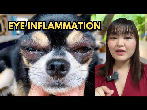
The symptoms of blepharitis are concentrated around the eyelids and the immediate periocular area, the skin and tissues surrounding the eye.
- Visible swelling and redness of one or both eyelids, particularly along the eyelid margin
- Crusting or scaling along the eyelid edge, which may be dry and flaky or moist and adhered
- Hair loss around the eye, with patchy or generalised thinning of the periocular fur
- Discharge from the eye, ranging from clear and watery to thick, yellow, or greenish pus, depending on whether infection is involved
- Squinting or partial closure of the affected eye
- Persistent rubbing of the face against furniture, carpet, or the dog’s own paws
- Thickening or roughening of the eyelid skin in chronic cases
- Small lumps, pustules, or ulcerated areas along the eyelid margin in more severe presentations
The itching and irritation associated with blepharitis are often more prominent than outright pain in mild cases. A dog with blepharitis will frequently be observed rubbing its face, sometimes to the point of causing self-inflicted skin damage around the eye.
Causes of Blepharitis in Dogs
Bacterial Infections
Bacterial infection of the eyelid skin and glands is one of the most common causes of blepharitis in dogs. Staphylococcal bacteria are frequent culprits, infecting the hair follicles and meibomian glands of the eyelid and producing the pustules, crusting, and discharge characteristic of infectious blepharitis. Bacterial blepharitis often develops secondarily to skin disease elsewhere on the body, reflecting a broader skin infection rather than an isolated eyelid problem.
Fungal and Parasitic Causes
Fungal infections, including ringworm, can affect the periocular skin and eyelid margins. Demodectic mange, caused by Demodex mites that inhabit hair follicles, is a significant parasitic cause of blepharitis, particularly in young dogs or immunocompromised individuals. Sarcoptic mange can also cause intense periocular irritation and secondary eyelid inflammation. Identifying parasitic causes requires specific diagnostic testing, as the treatment differs substantially from that used for bacterial or allergic disease.
Allergies
Allergic blepharitis is extremely common and is frequently the underlying driver of chronic or recurrent eyelid inflammation. Environmental allergens, including pollen, dust, and mould, can trigger an allergic response in the eyelid tissue. Contact allergy, where the eyelid skin reacts to something it directly touches, such as certain grasses or grooming products, is another presentation. Dogs with atopic dermatitis, a chronic skin allergy condition, frequently develop blepharitis as part of their broader allergic disease. In these dogs, the eyelids are one of multiple affected areas, and the condition is managed as part of a comprehensive allergy treatment plan.
Autoimmune and Immune-Mediated Disease
Several autoimmune conditions can produce blepharitis as either a primary or secondary feature. Pemphigus foliaceus, a condition where the immune system attacks the skin’s own structures, can affect the eyelid skin and cause crusting, erosion, and hair loss. Uveodermatological syndrome, a condition seen particularly in certain breeds including Akitas and Siberian Huskies, involves immune-mediated damage to both the eye and the pigmented skin around it. These immune-mediated causes require specific diagnosis and long-term immunosuppressive management.
Tumours and Eyelid Growths
Meibomian gland tumours, which are common in older dogs, can cause localised blepharitis by disrupting normal gland function and creating a focus of chronic inflammation. Other eyelid masses, including chalazia, which are blocked and inflamed meibomian glands, and histiocytomas, can produce similar localised swelling and irritation. In dogs with jaundice or metabolic disease affecting the liver, periocular skin changes can also occur. Our guide on Bile Duct Obstruction in Dogs covers how hepatic disease can produce systemic signs, including skin and ocular changes.
Related Videos


How Blepharitis Develops
The development of blepharitis follows a progression that begins with a trigger and, if unchecked, leads to chronic tissue disruption.
The initial trigger, whether infection, allergy, mite infestation, or immune response, causes inflammation in the eyelid skin or glands. This inflammation leads to gland dysfunction, particularly in the meibomian glands, whose oily secretion becomes altered or blocked. The altered secretion and inflamed tissue create an environment where bacteria can multiply more easily, and secondary infection often develops even in cases that began as purely allergic or immune-mediated.
Chronic inflammation thickens the eyelid tissue, distorts the eyelid margin, and can cause the eyelid to roll slightly inward or outward, bringing the abnormal lid margin into contact with the corneal surface. This contact creates a new source of corneal irritation and, over time, can cause corneal ulceration. What began as eyelid disease has now become a threat to the eye itself.
This progression is the reason early treatment matters. Blepharitis that is addressed before the eyelid structure is chronically distorted is far easier to manage and far less likely to cause secondary corneal damage.
Types of Blepharitis in Dogs
Blepharitis is categorised broadly by its underlying cause, which determines the treatment approach.
Infectious blepharitis involves bacterial, fungal, or parasitic organisms directly infecting the eyelid tissue. It often produces pustules, discharge, and crusting along the eyelid margin and responds to appropriate antimicrobial treatment once the causative organism is identified.
Allergic blepharitis is driven by the immune system’s response to environmental or contact allergens. It tends to be bilateral, affects multiple areas of the face in dogs with atopic disease, and is characterised by intense itching rather than pain. Management focuses on allergen avoidance and anti-inflammatory treatment.
Parasitic blepharitis caused by Demodex or Sarcoptes mites requires specific antiparasitic treatment and may need systemic therapy in generalised cases. Mite-associated blepharitis often occurs alongside skin disease elsewhere.
Immune-mediated blepharitis from autoimmune conditions requires immunosuppressive therapy and careful monitoring. These cases are typically more severe and more challenging to manage than infectious or allergic forms.
Diagnosis of Blepharitis in Dogs
Physical Examination
The veterinarian will examine the eyelids, eyelid margins, and periocular skin closely, assessing the character and distribution of swelling, crusting, hair loss, and discharge. The pattern of involvement, whether it is localised or diffuse, unilateral or bilateral, helps narrow the differential diagnosis.
Skin and Eyelid Tests
Skin cytology, where cells from the affected area are collected and examined microscopically, can identify bacteria, fungi, or inflammatory cell patterns. Skin scraping allows identification of mites. These tests are quick, minimally invasive, and provide information that directly guides treatment choices.
Allergy Testing
Where allergic blepharitis is suspected, particularly in dogs with a broader history of atopic disease, allergy testing through intradermal skin testing or serum allergy panels may be recommended to identify specific triggers. This information guides both avoidance strategies and allergen-specific immunotherapy if indicated.
Biopsy
In cases that are not responding to initial treatment, are producing an unusual clinical picture, or where an immune-mediated or neoplastic cause is suspected, a small biopsy of the affected eyelid tissue may be taken for histopathological examination. This provides definitive information about the type of inflammation present and guides specialist management.
Treatment of Blepharitis in Dogs
Warm Compresses and Eyelid Cleaning
Gentle cleaning of the eyelid margins with a warm, damp cloth removes crusting, discharge, and accumulated debris that perpetuates gland blockage and bacterial colonisation. Warm compresses applied for several minutes help to soften and express blocked meibomian gland secretions. These measures are supportive and practical and can be performed at home as directed by a veterinarian. They do not replace medical treatment but significantly support it.
Antibiotics and Antifungals
Bacterial blepharitis is treated with topical antibiotic ointments or drops applied to the eyelid margin and, in more extensive cases, with oral antibiotics. Treatment courses must be completed in full, even when symptoms appear to resolve early. Fungal blepharitis requires antifungal medication, and parasitic forms require appropriate antiparasitic treatment. The choice of medication depends on what the diagnostic testing has identified.
Anti-Inflammatory Medication
Where inflammation is prominent and infection has been controlled or ruled out, topical corticosteroid preparations may be used to reduce eyelid swelling and discomfort. In immune-mediated cases, systemic immunosuppressive treatment, including corticosteroids or other agents, may be required long-term. Anti-inflammatory treatment addresses the symptom. The underlying cause must be treated simultaneously.
Treating the Underlying Disease
This is the principle that determines long-term outcomes. Blepharitis that recurs repeatedly despite symptomatic treatment almost always has an underlying cause that is not being adequately addressed. Identifying and managing that cause, whether it is atopic disease, a systemic infection, an autoimmune condition, or a meibomian gland tumour, is what prevents the chronic cycle of inflammation, treatment, and recurrence.
Related Products


Long-Term Management
For many dogs, blepharitis is a condition that is controlled rather than cured. Dogs with underlying atopic disease, chronic autoimmune conditions, or recurrent meibomian gland dysfunction require ongoing management as a routine part of their care.
Long-term management typically involves periodic eyelid cleaning, maintenance anti-inflammatory treatment during flares, regular veterinary monitoring to catch early signs of recurrence or corneal involvement, and consistent management of the underlying disease. Owners who understand this and commit to the routine tend to achieve significantly better outcomes than those who treat each flare as an isolated event.
Complications if Left Untreated
Blepharitis that is inadequately managed or left untreated creates a progression of increasingly serious problems.
- Eyelid scarring from chronic inflammation distorts the normal eyelid structure and can cause permanent changes to the eyelid margin position
- Entropion, where the scarred eyelid rolls inward, and the lid margin or eyelashes contact the corneal surface continuously
- Corneal ulceration from the direct trauma of abnormal eyelid contact, which is painful and can threaten vision
- Corneal scarring from repeated superficial ulceration, causing permanent reduction in visual clarity
- Secondary deep eye infection if corneal ulcers become infected
The eye is vulnerable to what happens to the structures immediately around it. Eyelid disease that appears confined to the skin has a direct pathway to causing serious eye damage when left unmanaged.
Is Blepharitis Painful in Dogs?
Yes, though the quality and intensity of discomfort vary with the cause and severity.
Allergic blepharitis tends to cause intense itching, which drives the rubbing and pawing behaviour that is often the most visible sign. Dogs with allergic eyelid disease will rub their faces persistently, sometimes causing self-trauma that worsens the skin condition.
Infectious and immune-mediated blepharitis produce a combination of itching and pain, particularly when pustules, ulceration, or deep inflammation are involved. Dogs with severe infectious blepharitis may be reluctant to have the area touched and may show squinting and guarded behaviour more typically associated with ocular pain.
When blepharitis has progressed to cause corneal ulceration, the pain becomes significant and immediate. At this stage, the dog is experiencing two sources of discomfort simultaneously: the eyelid inflammation and the corneal wound, and veterinary attention is urgent.
Blepharitis vs Conjunctivitis: Understanding the Difference
These two conditions are frequently confused because they both involve the eye area and both produce redness and discharge. The distinction matters because the treatment approaches differ.
Blepharitis is inflammation of the eyelids themselves, the skin, glands, and tissue of the eyelid structure. The redness and swelling are in the lids. The crusting forms on the eyelid margins.
Conjunctivitis is inflammation of the conjunctiva, the thin, moist membrane lining the inner eyelids and covering the white of the eye. The redness is on the inner surface of the eyelid and the visible white of the eye. Discharge originates from the conjunctival surface.
Both conditions can occur simultaneously, and blepharitis frequently causes secondary conjunctivitis because the abnormal eyelid tissue directly contacts the conjunctival surface. However, treating only the conjunctivitis while blepharitis continues untreated is incomplete management.
Home Care vs Medical Treatment
Certain supportive measures can and should be performed at home under veterinary guidance.
Warm compresses and gentle eyelid margin cleaning with a clean, damp cloth are safe and beneficial. Removing crusting and softening blocked gland secretions supports healing and reduces bacterial load on the eyelid surface. These measures should be performed as directed by your veterinarian.
What should not be done at home without specific veterinary instruction is applying any medication to the eyelid or eye. Human antibiotic creams, corticosteroid preparations, antifungal creams, and over-the-counter eye drops are not safe for use in dogs without veterinary guidance. Some cause direct harm to the eye surface. Others mask symptoms while the underlying infection worsens. When in doubt about whether something is safe to apply, do not apply it. Contact your veterinarian first.
When to See a Vet
Seek veterinary assessment promptly if your dog shows:
- Persistent swelling or redness of one or both eyelids lasting more than a day or two
- Crusting, scaling, or discharge along the eyelid margin that is recurring or worsening
- Hair loss around the eyes
- Squinting, pawing at the eye, or rubbing the face repeatedly
- Any change in the appearance of the cornea or eye surface alongside eyelid symptoms
- Signs of pain when the area around the eye is touched
Early assessment leads to earlier diagnosis of the underlying cause and prevents the progression from eyelid disease to corneal complications.


