

Sometimes the problem is not the eye. It is the eyelashes.
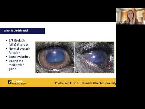
Distichiasis is a condition in which extra eyelashes grow from abnormal locations along the eyelid margin, specifically from the openings of the meibomian glands that sit at the inner edge of the lid. These additional lashes grow inward or at angles that bring them into direct contact with the surface of the eye. Every blink, every moment of eye movement, the aberrant lashes drag across the cornea.
In mild cases, this produces low-grade irritation that a dog may barely show. In more significant cases, it causes persistent pain, corneal ulceration, and, if left untreated, vision damage. The condition is chronic, often genetic, and does not resolve on its own.
What Is Distichiasis in Dogs?
The eyelid margin contains a row of specialised oil-producing glands called meibomian glands. These glands open onto the inner edge of the lid and contribute to the tear film that lubricates and protects the eye surface. In dogs with distichiasis, hair follicles develop within or adjacent to these gland openings, producing eyelashes that emerge from positions where no lashes should grow.
The aberrant lashes vary in character between affected dogs. In some dogs, they are soft and fine, causing minimal mechanical irritation. In others, they are stiff and coarse, producing significant abrasive contact with the corneal surface with every blink. The number of aberrant lashes can range from one or two to multiple lashes along one or both lids.
The condition is distinct from trichiasis, where normally positioned facial hair is misdirected toward the eye, and from ectopic cilia, where a single hair grows through the conjunctival surface of the lid directly into contact with the cornea. These three conditions produce similar symptoms through different mechanisms and are differentiated on careful examination of the lid margin.
Symptoms of Distichiasis in Dogs
The clinical signs of distichiasis reflect the degree of corneal irritation the aberrant lashes are producing.
- Excessive tearing and wet staining of the fur below the inner corner of the eye
- Squinting, increased blinking, or keeping one or both eyes partially closed
- Visible redness of the conjunctiva and the sclera around the eye
- Pawing at or rubbing the affected eye against furniture or the floor
- Mucoid or mucopurulent discharge from the eye
- Conjunctivitis that recurs or does not resolve with standard treatment
- Corneal cloudiness, irregularity, or visible ulceration in more advanced or untreated cases
Some dogs with soft, fine distichia show no clinical signs at all. The lashes are present and detectable on careful slit-lamp examination, but do not produce enough friction on the corneal surface to trigger a visible inflammatory response. These cases may be monitored rather than treated immediately. However, the absence of obvious signs does not mean the lashes are harmless, and monitoring must continue because the corneal surface remains at ongoing risk.
Causes of Distichiasis in Dogs
Genetic Predisposition
Distichiasis is primarily a genetic condition. The abnormal development of hair follicles within the meibomian gland architecture is an inherited trait in many affected breeds. Dogs from affected lines carry the predisposition regardless of whether their individual cases produce significant clinical signs.
Breed Predisposition
Certain breeds are significantly overrepresented in distichiasis diagnoses. Shih Tzus, Cocker Spaniels, English Bulldogs, Golden Retrievers, Boxers, Poodles, and Dachshunds are among the breeds most commonly affected. In these breeds, the condition is so prevalent that ophthalmoscopic examination of the lid margin should be a routine part of health screening.
Chronic Glandular Inflammation
In some cases, chronic inflammation of the meibomian glands may stimulate the development of aberrant hair follicles within the gland tissue. This represents an acquired rather than purely congenital mechanism and may explain the occasional presentation of distichiasis in breeds or individuals without a strong genetic predisposition.
Related Videos


How Distichiasis Affects the Eye
The cornea is the transparent tissue at the front of the eye. It is densely innervated and highly sensitive to mechanical contact. When distichia rub against the corneal surface, even soft lashes produce friction that disrupts the normal tear film distribution and creates microtrauma to the epithelial cells of the corneal surface.
With repeated contact over time, this microtrauma accumulates. The corneal epithelium becomes irregularly disrupted. Superficial corneal ulcers develop where the lash contact is most sustained and most direct. Corneal ulcers are painful, prone to secondary bacterial infection, and if they progress to deeper layers of the cornea, they can cause significant and permanent vision damage.
The most serious complication of untreated distichiasis is the development of corneal scarring, where the corneal tissue that heals following ulceration becomes opaque rather than transparent, reducing or blocking the passage of light to the retina. This corneal fibrosis is irreversible, which is why eye ulcer in dogs represents one of the most important complications that timely treatment of distichiasis directly prevents.
How Veterinarians Diagnose Distichiasis in Dogs
Physical Eye Examination
The lid margins are examined carefully under magnification, typically using a slit-lamp biomicroscope or a bright focused light source. The meibomian gland openings are inspected for aberrant lash emergence. This examination requires the dog to be cooperative and may need sedation in anxious or painful animals to allow thorough assessment.
Fluorescein Stain Testing
Fluorescein dye is applied to the eye surface to identify any corneal ulceration. The dye adheres to areas of disrupted corneal epithelium and fluoresces under blue light, clearly delineating the location and extent of any ulceration. This test is performed at the time of diagnosis and at follow-up appointments to monitor the corneal surface.
Assessment of Severity
The character of the distichia, whether soft and fine or coarse and stiff, their number and location, and the degree of corneal involvement together determine the appropriate treatment approach.
Treatment for Distichiasis in Dogs
Treatment is matched to the severity of the condition and the degree of clinical impact.
Lubricating Eye Drops and Ointments (Mild Cases)
In dogs with soft, fine distichia producing minimal corneal irritation, lubricating eye drops or ointments applied regularly can reduce friction between the lashes and the corneal surface and manage symptoms without addressing the lashes directly. This is a palliative approach appropriate only for genuinely mild cases and requires regular monitoring to ensure the corneal surface remains healthy.
Epilation (Temporary)
Manual plucking of the aberrant lashes under sedation provides immediate relief of the mechanical irritation. The lashes are removed at the surface, but the hair follicles within the gland tissue remain intact. Regrowth of the distichia typically occurs within four to six weeks, and repeated epilation is required to maintain relief. This is used as a temporary measure or as a management strategy for dogs that are not candidates for more definitive treatment.
Cryotherapy (Permanent Treatment)
Cryotherapy applies controlled freezing to the meibomian gland tissue at the sites of aberrant follicle development. The extreme cold destroys the follicular cells responsible for producing the distichia without requiring incision into the eyelid. It is one of the most effective treatments for permanent resolution of distichiasis and has a high success rate when performed by a veterinary ophthalmologist. A degree of eyelid depigmentation at the treated site is an expected and typically cosmetically acceptable side effect.
Electrolysis and Electroepilation
Fine needle electroepilation applies electrical current directly to individual aberrant follicles to destroy them. This technique is precise and effective for individual or small numbers of distichia but is less practical when large numbers of lashes require treatment across multiple sites.
Surgical Treatment
Surgical excision of the meibomian gland tissue containing the aberrant follicles is used in cases not amenable to cryotherapy or electrolysis. This approach removes the tissue source of the distichia but requires careful surgical technique to avoid damaging the lid margin architecture and compromising normal meibomian gland function.
Related Products

Distichiasis, Trichiasis, and Ectopic Cilia
| Feature | Distichiasis | Trichiasis | Ectopic Cilia |
|---|---|---|---|
| Origin of hair | Meibomian gland openings | Normal facial or nasal fold hair | Single hair through conjunctival surface |
| Direction of growth | Toward or along corneal surface | Redirected toward eye from normal position | Directly into cornea from inside lid |
| Severity | Mild to moderate typically | Varies with breed conformation | Often severe and acutely painful |
| Treatment | Cryotherapy, epilation, surgery | Manage facial folds, hair removal | Surgical excision of follicle |
Understanding these distinctions matters because each condition requires a different treatment approach and carries different implications for the dog’s prognosis. Misidentifying the specific condition risks providing treatment that addresses the wrong problem.
Is Distichiasis Painful?
This depends on the character of the lashes and the degree of corneal involvement. Dogs with soft, fine distichia and no corneal ulceration may show minimal signs of discomfort and appear outwardly normal despite the lashes being identifiable on examination.
Dogs with coarse, stiff lashes or with lashes making sustained direct contact with the corneal surface experience real and significant discomfort. The cornea is one of the most sensitive tissues in the body. Chronic mechanical irritation produces the equivalent of sustained corneal abrasion, and the associated pain affects the dog’s normal behaviour, reducing their willingness to open the eye fully and producing the persistent squinting and pawing that characterise more significant cases.
Prognosis and Long-Term Management
With appropriate treatment, the prognosis for distichiasis is good. Most dogs achieve significant or complete relief of symptoms following cryotherapy or other permanent follicle destruction procedures. Regular monitoring of the corneal surface remains important even after treatment because recurrence of distichia from residual follicular tissue is possible, and the cornea requires assessment to confirm continued health.
Dogs that have sustained corneal scarring before treatment will retain those scars regardless of treatment success. The opacity and any associated vision impairment from pre-existing scarring are not reversed by treating the distichiasis, which reinforces the importance of early diagnosis and treatment before corneal damage accumulates.
When to See a Veterinarian
Contact your veterinarian promptly if your dog shows any of the following:
- Persistent or increasing tearing from one or both eyes
- Squinting, excessive blinking, or difficulty keeping the eye open
- Visible redness of the eye or surrounding tissue
- Repeated pawing at the eye or rubbing the face
- Mucoid or purulent eye discharge
- Any visible change in the clarity or surface of the cornea
Eye conditions in dogs do not resolve on their own in the majority of cases, and distichiasis left without assessment allows corneal damage to accumulate. Early examination and treatment prevent the progression from manageable irritation to corneal ulceration and scarring.
Early veterinary assessment is part of responsible dog ownership, and the full range of conditions that can affect your dog is covered comprehensively in the VOSD dog medical conditions library.


