

The mouth is one of the last places most pet parents think to examine regularly, and that delay is one of the main reasons oral cancer in dogs is so often diagnosed at an advanced stage. Mouth cancer in dogs refers to malignant tumours arising from the tissues of the oral cavity: the gums, tongue, lips, hard and soft palate, and the underlying jawbone. The most common types are oral melanoma, squamous cell carcinoma (SCC), fibrosarcoma, and acanthomatous ameloblastoma, each with distinct behaviour, aggressiveness, and response to treatment. What they share is this: dogs whose tumours are found and treated early consistently do better than those whose tumours are found in crisis. Regular oral examination, at home and by a veterinarian, is one of the most practical things a pet parent can do.
Quick Overview: Key Facts at a Glance
| Feature | Details |
| Condition | Oral cancer (malignant tumours of the mouth) |
| Common tumour types | Oral melanoma, squamous cell carcinoma, fibrosarcoma, acanthomatous ameloblastoma |
| Most common type | Oral melanoma accounts for the majority of malignant oral tumours in dogs |
| Affected age group | Primarily middle-aged to senior dogs; typically over 8 years |
| Key symptoms | Oral mass or swelling, bad breath, drooling, bleeding, difficulty eating |
| Primary treatment | Surgical removal, radiation, and immunotherapy as adjuncts, depending on the type |
| Prognosis (localised, treated) | Varies significantly by tumour type, stage, and treatment approach |
| Most important factor | Early detection before lymph node or bone involvement |
What Is Dog Mouth Cancer?
Oral tumours develop when cells within the mouth’s soft or hard tissues begin to replicate abnormally, forming a mass that invades surrounding structures rather than remaining contained. In dogs, the oral cavity is the fourth most common site for cancer, a higher incidence than many pet parents realise.
Not all oral masses in dogs are malignant. Some are benign growths, epulides, fibromas, papillomas, that can be removed without the staging or adjuvant treatment that malignant tumours require. However, benign and malignant oral masses can appear clinically similar, and the appearance of a growth alone is never sufficient to determine malignancy. Tissue examination is always required. For an overview of the full range of oral growths, including how benign and malignant masses differ in presentation and management, oral masses in dogs covers this in detail.
The oral cavity is a challenging site for cancer management because tumours here can invade the jawbone, involve regional lymph nodes early, and interfere with essential functions, eating, drinking, and swallowing, even before they have metastasised distantly.
Types of Mouth Cancer in Dogs
Oral Melanoma
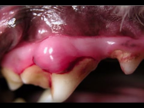
Oral melanoma is the most common malignant oral tumour in dogs, and one of the most aggressive. It arises from melanocytes, pigment-producing cells, within the oral mucosa, gums, or lips. Oral melanomas are locally invasive, invade bone early, and metastasise to regional lymph nodes and lungs at a high rate – in many cases, metastasis has occurred by the time of diagnosis, even when the primary tumour appears small. Darkly pigmented masses on the gums are the most recognised presentation, but oral melanoma can also be amelanotic (non-pigmented), appearing pink or pale, which delays recognition. Staging and treatment are available for mouth cancer (melanocytic) in dogs.
Squamous Cell Carcinoma (SCC)
Squamous cell carcinoma of the gingiva (gum tissue) arises from the squamous epithelial cells lining the oral mucosa. It is locally very invasive, bone invasion is common, but metastasises to distant sites less frequently than melanoma, particularly in its rostral (front of the mouth) location. Tonsillar SCC, by contrast, carries a very poor prognosis with high metastatic rates. Gingival SCC often appears as a proliferative, ulcerated mass associated with the gum line, and may initially be mistaken for periodontal disease. A specific treatment approach is also available on VOSD for mouth cancer, gingiva squamous cell carcinoma in dogs.
Fibrosarcoma
Fibrosarcoma arises from the connective tissue (fibroblasts) of the oral cavity, commonly affecting the gums of the upper jaw and the hard palate. It is locally aggressive with high recurrence rates after surgery, but metastasises less readily than melanoma. Fibrosarcomas are often firm masses that invade the underlying bone, and achieving adequate surgical margins is technically challenging given the location.
Acanthomatous Ameloblastoma
Formerly called acanthomatous epulis, this tumour arises from dental tissue and is locally aggressive; it invades bone significantly, but does not metastasise. Despite being considered a locally malignant rather than systemically malignant tumour, it requires aggressive local treatment (typically surgical jaw resection) to prevent recurrence. It is most common in middle-aged dogs and is particularly associated with certain breeds, including the Shetland Sheepdog. More Details available for mouth cancer and ameloblastoma in dogs.
Related Videos


Symptoms of Mouth Cancer in Dogs
Oral tumours can grow for some time before producing obvious symptoms, particularly on the palate or the back of the mouth, where routine visibility is limited. The first sign is often a behavioural change that the owner interprets as something else: the dog eating more slowly, being reluctant to chew harder food, or pawing at their face.
- Visible lump, mass, or swelling in or around the mouth, on the gums, under the tongue, on the palate, or producing facial asymmetry from below
- Halitosis (bad breath), more severe or qualitatively different from typical dental odour; often described as unusually foul or necrotic-smelling
- Drooling, increased saliva production, sometimes blood-tinged; the dog may drip saliva when resting
- Bleeding from the mouth, from the tumour surface, may be noticed on bedding, toys, or water bowls
- Loose or displaced teeth, bone invasion by the tumour disrupts tooth roots; a loose tooth without obvious periodontal disease is a red flag
- Difficulty eating or chewing, the dog chews on one side, drops food, refuses hard food, or eats more slowly and reluctantly than before
- Dysphagia (difficulty swallowing), in tumours involving the caudal oral cavity or pharynx
- Weight loss, secondary to reduced food intake from oral pain or mechanical obstruction
- Facial swelling or asymmetry, visible deformity of the face or jaw in cases with significant bone involvement
- Pawing at the face or mouth, a behavioural sign of oral discomfort, often precedes the owner identifying any visible cause
- Enlarged submandibular lymph nodes, palpable swelling under the jaw, may indicate regional metastasis
Any of these signs persisting for more than one to two weeks should prompt a veterinary oral examination rather than home monitoring.
Causes and Risk Factors
Oral cancer in dogs does not have a single identifiable cause. Like most cancers, it is understood as the product of multiple interacting factors:
Age – the large majority of cases, are diagnosed in dogs over eight years old. The accumulation of genetic mutations over time and the gradual decline of immune surveillance both contribute to age as the most consistent risk factor.
Breed and pigmentation – dogs with heavily pigmented oral mucosa (dark-gummed breeds) are at higher risk for oral melanoma. Cocker Spaniels, German Shepherds, Weimaraners, Golden Retrievers, and small breeds, including Miniature
Poodles – appear more frequently in oral melanoma case series. Flat-coated Retrievers have an elevated risk of oral fibrosarcoma. The Shetland Sheepdog is overrepresented in ameloblastoma cases.
Chronic oral inflammation – persistent gingivitis, periodontal disease, and chronic oral infection create sustained inflammatory microenvironments that promote abnormal cell proliferation. Regular dental care is not just about breath and tooth retention; it is a meaningful cancer-risk-reduction measure.
Environmental exposures – secondhand tobacco smoke, chemical exposure, and urban air pollution have been suggested as contributing factors in canine oral cancer epidemiology, though the strength of individual associations is not definitively established.
Sex – male dogs have a somewhat higher incidence of oral tumours than females, particularly melanoma.
How Veterinarians Diagnose Dog Mouth Cancer
Accurate diagnosis requires a structured process that identifies the tumour type, assesses local invasion, and stages systemic spread before any treatment decision is made.
Step 1
Oral examination: A thorough examination under sedation or anaesthesia, assessing all surfaces of the oral cavity including the palate, base of the tongue, and tonsillar recesses, areas not fully visible in an awake, moving dog. All accessible lymph nodes are palpated.
Step 2
Dental radiographs: Intraoral dental radiographs reveal bone involvement beneath the gum line; essential for characterising the extent of local invasion before surgery.
Step 3
Biopsy and histopathology: The definitive diagnostic step. A tissue sample is taken from the mass and submitted for histopathological analysis. Immunohistochemistry may be used to confirm melanocytic differentiation in amelanotic tumours. The diagnosis, grade, and margin assessment from the biopsy guide all subsequent decisions.
Step 4
CT scanning: Provides detailed three-dimensional assessment of bone invasion, extension into the nasal passages or orbit, and regional lymph node involvement. CT is considered standard of care for surgical planning of oral tumours and is increasingly available at veterinary referral centres.
Step 5
Lymph node assessment: Mandibular and retropharyngeal lymph nodes are aspirated for cytology; even clinically normal-sized lymph nodes can harbour metastatic cells, and formal lymph node staging influences both prognosis and treatment planning.
Step 6
Thoracic staging: Three-view thoracic radiographs assess for pulmonary metastasis; CT of the thorax provides greater sensitivity if available.
Treatment Options for Dog Mouth Cancer
Surgery is the primary treatment for most localised oral tumours. The type of surgery depends on the tumour’s size, location, and extent of bone invasion:
Mandibulectomy (partial or complete removal of the lower jaw) and maxillectomy (partial removal of the upper jaw) are the procedures most commonly required for oral tumours with bone involvement. These are significant surgeries, but dogs adapt to them far better than owners anticipate, most dogs are eating and maintaining quality of life within days to weeks of surgery, because their anatomy allows function without the full jaw structure.
Achieving adequate surgical margins, a border of normal tissue around the tumour, is the primary determinant of local recurrence. Incomplete margins significantly increase recurrence risk.
Radiation therapy is used as a primary treatment when surgery cannot achieve adequate margins, and as adjuvant therapy post-surgery in high-risk cases. It is the primary treatment modality for oral melanoma with nodal involvement, and effective for acanthomatous ameloblastoma when surgery is not feasible. Available at veterinary oncology referral centres; requires multiple anaesthetic sessions.
Immunotherapy (melanoma vaccine), a canine melanoma DNA vaccine (Oncept) is licensed for the treatment of oral melanoma in dogs and works by stimulating the immune system to target melanocyte-specific antigens. It is used after local disease control (surgery and/or radiation) to address micrometastatic disease. The vaccine has extended median survival times in dogs with oral melanoma in clinical studies and represents a meaningful advance in oral melanoma management.
Chemotherapy has limited efficacy as a primary treatment for oral melanoma and SCC, but is used as part of multimodal protocols for metastatic disease management.
Palliative care – for dogs where curative-intent treatment is not feasible, pain management (NSAIDs, opioids, gabapentin), nutritional support, and regular reassessment of quality of life are central. A dog eating comfortably, engaging with their environment, and free from pain is the benchmark.
Related Products


Life Expectancy of Dogs With Mouth Cancer
Survival varies significantly by tumour type, stage, and treatment approach. Honest benchmarks are more useful than vague reassurance:
Oral melanoma, untreated: Median survival of approximately 65 days from diagnosis.
Oral melanoma, surgery alone: Median survival of approximately 5–17 months depending on stage at surgery; Stage I (tumour under 2 cm, no nodal metastasis) has a significantly better outcome than Stage III.
Oral melanoma, surgery with radiation and melanoma vaccine: Median survival times extend further; some studies report median survival over 14 months with multimodal treatment, with a subset of dogs surviving well beyond two years.
Squamous cell carcinoma (rostral, surgically resected with clean margins): Median survival of one year or more; rostral SCC is considered the most favourable oral cancer for surgical outcomes.
Squamous cell carcinoma (tonsillar): Median survival is typically under 6 months, even with treatment, due to a high metastatic rate.
Fibrosarcoma: Median survival with surgery is approximately 10–12 months; high local recurrence rate even after resection.
Acanthomatous ameloblastoma (surgery with adequate margins): Excellent long-term prognosis; recurrence is low when margins are clean; does not metastasise.
Prognosis Factors
Several factors consistently influence outcomes across all oral tumour types:
Tumour size at diagnosis – smaller tumours at the time of treatment are associated with better outcomes across all types. Oral melanoma staging is based on tumour size: Stage I (under 2 cm), Stage II (2–4 cm), Stage III (over 4 cm or regional lymph node metastasis).
Location within the oral cavity, rostral (front of the mouth) tumours are more accessible, easier to resect with margins, and are generally associated with better outcomes than caudal (back of the mouth) tumours.
Bone invasion – tumours that have invaded the underlying jawbone require more extensive surgery, are more likely to have incomplete margins, and are associated with shorter disease-free intervals.
Lymph node and pulmonary metastasis are the single most significant prognostic factors. Confirmed metastasis at diagnosis changes the goal from curative to palliative-intent treatment.
Surgical margin status – histopathological confirmation of clean margins after surgery- is strongly associated with longer disease-free survival.
Dogs’ overall health and performance status – dogs with good organ function who can safely undergo surgery and recovery have more treatment options than dogs with concurrent systemic illness.
When to See a Veterinarian
Book a veterinary appointment within the week if you notice any visible growth or mass in your dog’s mouth, persistent bad breath that does not improve with dental hygiene, saliva or discharge that is blood-tinged, a loose tooth without obvious injury, facial asymmetry, or any change in eating behaviour, eating slower, dropping food, favouring one side, or refusing hard food. Do not wait to see if it resolves. Oral tumours do not shrink or disappear on their own, and the difference between finding a tumour at 1 cm and finding it at 3 cm with bone involvement is often the difference between a resectable and a non-resectable case.
Prevention and Oral Health Tips
While oral cancer cannot be fully prevented, its risk can be reduced, and its likelihood of early detection significantly improved:
- Regular home oral examinations, lift the lips, look at the gum line, check the roof of the mouth and under the tongue; monthly checks allow early identification of any new growth
- Routine dental care, brushing (or enzymatic dental products for dogs who resist brushing), professional veterinary dental cleaning, and management of periodontal disease reduce chronic oral inflammation
- Annual or biannual veterinary oral examination, particularly in dogs over six and in at-risk breeds; a thorough oral examination under sedation is part of every professional dental cleaning
- Prompt investigation of any oral mass, even one that appears benign; fine needle aspiration or biopsy is the only way to know with certainty
- Act on behavioural changes early; a dog eating differently is telling you something – oral pain is the most common reason, and it should be investigated
Conclusion
Oral cancer in dogs is more common than most people realise, and its outcomes are more influenced by timing than by any other single factor. A tumour found at Stage I and treated with surgery, appropriate adjuvant therapy, and proper staging has a meaningfully different outcome than the same tumour found at Stage III. The gap between those two presentations is often filled with months of symptoms that were present but not investigated. Routine oral examination, at home, at every vet visit, and formally under anaesthesia at annual dental assessments, is the practical, accessible tool that bridges that gap. Know what your dog’s mouth normally looks like, and act when something changes.


