

Not all vision problems begin with age. Some begin before a puppy ever opens its eyes.
Congenital eye defects are structural abnormalities that are present at birth or develop in the earliest weeks of a dog’s life, during the critical period when the eyes are still forming. They are not caused by something the dog encountered, caught, or was exposed to after birth. They are the result of disruptions that occurred during development, whether genetic, nutritional, or environmental in origin, before the puppy entered the world.
What makes congenital eye defects particularly important to understand is that they exist on an enormous spectrum. Some are mild, stable, and never cause the dog significant difficulty. Others are progressive, painful, and can ultimately lead to blindness if left unaddressed. The challenge for owners is that a puppy with a congenital eye defect may appear entirely normal in the early weeks of life, with no obvious signs of a problem until the condition has already advanced.
Early identification is the single most important factor in determining how well an affected dog does over the long term. And early identification begins with owners and breeders knowing what to look for and when to act.
What Are Congenital Eye Defects in Dogs?
Congenital eye defects are structural or functional abnormalities of the eye that are present at birth or emerge during early postnatal development. The word congenital refers specifically to the timing of origin, meaning the defect was established during the developmental process rather than acquired through injury or disease later in life.
Some congenital defects are immediately visible when a puppy’s eyes first open at around two weeks of age. Others are not detectable without a veterinary examination, and some only become apparent as the puppy grows and the eyes develop further. This variability means that a puppy that appears normal at birth can still carry a significant congenital eye condition that will not reveal itself until weeks or months later.
Congenital eye defects can affect any structure of the eye, including the eyelids, the cornea, the lens, the retina, the iris, and the overall size and shape of the eye itself. Each structure, when affected, produces its own characteristic pattern of signs and carries its own implications for vision and long-term health.
Types of Congenital Eye Defects in Dogs
Understanding the range of congenital eye defects that exist helps owners and breeders recognise what they may be dealing with and communicate meaningfully with their veterinarian.
Congenital cataracts involve clouding of the lens that is present from birth or develops in early puppyhood. Unlike age-related cataracts, which develop in older dogs, congenital cataracts are caused by genetic factors or developmental disruptions during lens formation. They can affect one or both eyes and vary in severity from small opacities that do not significantly impair vision to complete lens clouding that causes blindness.
Congenital glaucoma is a condition in which drainage of the fluid inside the eye is structurally impaired from birth, leading to elevated intraocular pressure. This pressure causes pain, progressive damage to the optic nerve, and eventually blindness if not managed. It is less common than acquired glaucoma but carries serious consequences when present.
Retinal dysplasia is a developmental abnormality in which the retinal layers fail to form correctly, resulting in folds, rosettes, or detachments within the retinal tissue. Mild forms may cause minimal vision impairment, while severe forms involving complete retinal detachment result in blindness. It is seen with particular frequency in Labrador Retrievers, Rottweilers, and English Springer Spaniels.
Coloboma is a gap or notch in one of the eye’s structures, most commonly the iris, choroid, or optic disc, resulting from incomplete closure during fetal development. The severity depends entirely on which structure is affected and how large the gap is. Colobomas of the iris are often visible as an irregular pupil shape. Those affecting the retina or optic disc can significantly impair vision.
Dermoids are patches of skin-like tissue that form on the cornea or conjunctiva. They contain skin structures, including hair follicles, and the hairs that grow from them cause chronic irritation of the eye surface. Dermoids are typically visible and almost always require surgical removal.
Microphthalmia is a condition in which one or both eyes are abnormally small due to incomplete ocular development. Affected eyes may have limited or absent visual function depending on the degree of underdevelopment. Microphthalmia is associated with several genetic mutations and is seen in certain breeds predisposed to merle coat genetics.
Iris cysts are fluid-filled structures that form within or behind the iris. While some iris cysts are benign and do not affect vision or cause discomfort, others can grow large enough to interfere with vision or cause secondary complications, including elevated intraocular pressure.
Symptoms of Congenital Eye Defects in Dogs
The symptoms of congenital eye defects vary significantly depending on the type and severity of the defect, but there are common patterns that should prompt owners to seek veterinary assessment without delay.
Cloudy or hazy eyes in a young puppy are never normal. Cloudiness of the lens suggests cataracts. Cloudiness of the cornea may indicate corneal abnormalities or elevated intraocular pressure from congenital glaucoma.
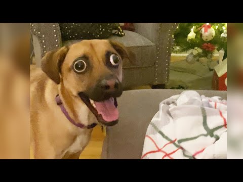
Visible abnormalities in eye structure, including an irregular pupil shape, an unusually small eye, visible tissue on the corneal surface, or asymmetry between the two eyes, are direct indications of a structural defect that requires examination.
Signs of vision impairment, including difficulty navigating familiar environments, bumping into objects, reluctance to move in low light, or failure to track moving objects normally, suggest that the defect is affecting visual function.
Redness, discharge, and signs of pain, including squinting, pawing at the face, or sensitivity to light, indicate that the defect is causing active irritation or inflammation. These signs require prompt attention as they may indicate secondary complications such as corneal damage or elevated pressure.
Enlarged eye in one or both eyes in a young dog is a concerning sign associated with congenital glaucoma, where elevated intraocular pressure causes the eye to expand. This is a condition requiring urgent veterinary assessment.
Related Videos


Causes of Congenital Eye Defects in Dogs
Genetic Inheritance
The majority of congenital eye defects in dogs have a genetic basis. They are caused by inherited mutations that disrupt normal eye development at specific stages of fetal or early postnatal growth. In many breeds, specific genetic mutations linked to particular eye conditions have been identified, and testing for these mutations is available through veterinary genetic screening.
The genetic transmission of congenital eye defects follows different inheritance patterns depending on the specific condition. Some defects are autosomal recessive, meaning both parents must carry the gene for a puppy to be affected. Others are autosomal dominant, meaning a single copy of the mutation is sufficient to cause the condition.
Developmental Disruptions During Pregnancy
Not every congenital eye defect is directly inherited. Disruptions to normal eye development during pregnancy can produce structural abnormalities even in puppies without a genetic predisposition.
Nutritional deficiencies in the mother during pregnancy, particularly deficiencies in vitamins essential for fetal development, can interfere with normal ocular formation.
Infections during pregnancy, particularly certain viral infections that cross the placental barrier, can disrupt the developmental timeline of the eyes and produce structural abnormalities in the puppies.
Exposure to toxins or certain medications during pregnancy can also interfere with normal fetal eye development, producing congenital defects that are environmental rather than genetic in origin.
Understanding the cause of a congenital defect matters because it has direct implications for breeding decisions and for the management of the affected dog.
Breeds at Higher Risk
Congenital and inherited eye defects have been documented across many breeds, but certain breeds carry a significantly elevated risk due to well-established genetic predispositions.
The Rough Collie and Border Collie are strongly associated with Collie Eye Anomaly, a genetic condition affecting the development of the choroid and retina that ranges from mild and non-progressive to severe with retinal detachment.
The Siberian Husky and Alaskan Malamute are predisposed to hereditary cataracts and certain corneal conditions. Labrador Retrievers and Golden Retrievers carry a higher incidence of retinal dysplasia and progressive retinal atrophy. Cocker Spaniels are predisposed to both congenital cataracts and glaucoma.
Breeds with merle coat genetics, including the Australian Shepherd, Dachshund, and Great Dane, are at significantly elevated risk of microphthalmia and other serious developmental eye abnormalities, particularly when two merle-carrying dogs are bred together.
Owners of these breeds should ensure that puppies receive a thorough ophthalmic examination from a veterinarian experienced in canine eye conditions, ideally before eight weeks of age and repeated during the first year of life.
Diagnosis of Congenital Eye Defects in Dogs
Diagnosis of congenital eye defects requires a systematic ophthalmic examination combined with targeted testing to assess each component of the eye and identify any structural or functional abnormality.
Ophthalmoscope examination allows the veterinarian to visualise the internal structures of the eye, including the retina, optic disc, lens, and vitreous. This is essential for identifying retinal dysplasia, colobomas, cataracts, and other internal defects that are not visible on the surface of the eye.
The Schirmer tear test assesses tear production to ensure that the eye surface is adequately lubricated and that dry eye is not contributing to any surface abnormalities.
Tonometry measures intraocular pressure and is essential for identifying congenital glaucoma, where elevated pressure is the defining diagnostic feature and the driver of progressive optic nerve damage.
Fluorescein staining of the cornea identifies any surface ulceration, which can develop secondary to congenital defects that disrupt the corneal surface, including dermoids and certain structural abnormalities of the eyelids.
Ocular ultrasound is used when internal structures cannot be adequately visualised due to lens opacity or other obstructions. It is particularly useful for assessing retinal position and identifying lens luxation or other posterior segment abnormalities.
Genetic testing is available for a growing number of breed-specific congenital eye conditions. In predisposed breeds, genetic testing of breeding animals is a responsible and important step in reducing the incidence of heritable eye disease in future generations.
Treatment of Congenital Eye Defects in Dogs
The treatment of congenital eye defects depends entirely on the type of defect, the structures affected, and the degree to which the condition is impairing the dog’s comfort and vision.
Medical Management
Some congenital defects, particularly those causing surface irritation or secondary inflammation without structural compromise requiring surgery, are managed medically.
Lubricating eye drops and artificial tear solutions are used to protect the corneal surface in dogs where the defect disrupts normal tear distribution or exposes the eye surface to increased friction and dryness.
Topical anti-inflammatory or antibiotic drops may be prescribed when secondary inflammation or infection is present alongside the primary structural defect.
For dogs with iris cysts that are not causing significant complications, regular monitoring rather than active treatment is the appropriate management approach, with intervention reserved for cases where the cysts grow large enough to affect vision or intraocular pressure.
Surgical Management
Several congenital eye conditions require surgical correction to restore function or prevent progressive damage.
Cataract surgery involving removal of the clouded lens and, in many cases, implantation of an artificial lens, is the definitive treatment for congenital cataracts that are impairing vision. Early surgical intervention in young dogs with significant cataracts is important because prolonged visual deprivation during the critical period of visual development can itself cause permanent changes in the visual processing pathways.
Surgical removal of dermoids is almost always indicated, as the hairs growing from these lesions cause chronic corneal irritation that will not resolve without removal of the abnormal tissue.
Glaucoma management in congenital glaucoma may involve both medical treatment to reduce intraocular pressure and surgical procedures to improve fluid drainage from the eye. Early and aggressive management is essential to preserve any remaining vision.
Conditions Where Management Replaces Cure
For conditions such as retinal dysplasia, colobomas of the retina or optic disc, and microphthalmia, there is no surgical correction that restores the missing or malformed tissue. Management focuses on protecting the dog from environments where its visual impairment creates a safety risk, monitoring for secondary complications, and ensuring the dog’s overall quality of life is maintained despite the structural limitation.
Prognosis and Long-Term Outlook
The prognosis for dogs with congenital eye defects varies as widely as the conditions themselves. Making a meaningful statement about prognosis requires knowing the specific defect, the degree of structural involvement, and whether the condition is stable or progressive.
Dogs with mild congenital cataracts affecting only part of the lens, or with focal retinal dysplasia without detachment, or with small benign iris cysts often live functionally normal lives with minimal or no impact on their day-to-day vision.
Dogs with progressive conditions such as advanced hereditary cataracts, severe retinal dysplasia with detachment, or congenital glaucoma face a more challenging outlook that requires active management and realistic expectations. These dogs can often adapt remarkably well to significant visual impairment, particularly when the vision loss is gradual, but they require an environment that accounts for their limitations and an owner who understands what those limitations mean in practical terms.
The key factor in long-term outcome across all types of congenital eye defect is how early the condition is identified and how consistently it is managed from that point forward.
Complications If Left Untreated
Congenital eye defects that are not identified and managed appropriately carry a predictable risk of progressive complications that significantly worsen the dog’s situation over time.
Vision loss and blindness are the most serious potential outcomes. Conditions that are treatable or manageable in their early stages, such as congenital cataracts or congenital glaucoma, can lead to permanent blindness when left unaddressed for too long. The window for effective intervention is often narrow.
Chronic pain becomes a factor when structural defects lead to ongoing corneal irritation, elevated intraocular pressure, or surface ulceration. A dog with an unmanaged dermoid, advanced congenital glaucoma, or chronic corneal trauma from a structural eyelid abnormality is living with ongoing discomfort that is entirely preventable with appropriate care.
Secondary infections develop more readily in eyes with compromised structural integrity, as the normal protective barriers of the eye are disrupted. These infections add further damage to a surface that is already vulnerable.
Irreversible structural damage to the retina, optic nerve, or cornea accumulates with time in conditions where elevated pressure or chronic inflammation is not managed. This damage cannot be reversed once it has occurred.
Common Genetic Eye Disorders in Dogs
Several conditions deserve specific mention because of how frequently they appear in clinical practice and how significant their impact can be on affected dogs.
Collie Eye Anomaly is one of the most prevalent inherited eye conditions in herding breeds. It involves abnormal development of the choroid, the vascular layer beneath the retina, and ranges from mild choroidal hypoplasia to severe disease with retinal detachment and haemorrhage. Genetic testing is available and widely used in responsible breeding programmes for affected breeds.
Progressive Retinal Atrophy (PRA) is a group of inherited conditions in which the photoreceptor cells of the retina degenerate progressively over time, leading to night blindness first and eventually complete blindness. It affects numerous breeds and is among the most well-studied inherited eye diseases in dogs.
Corneal dystrophy, discussed in detail in VOSD’s guide to inherited corneal conditions, is a genetic disorder affecting the clarity of the cornea through lipid or fluid accumulation within the corneal layers.
Lens luxation, though sometimes acquired, has a strong inherited component in certain breeds, including the Tibetan Terrier, Jack Russell Terrier, and Border Collie. It involves displacement of the lens from its normal position, which can cause secondary glaucoma and requires urgent management when it occurs.
When secondary inflammation develops in eyes affected by congenital defects, the resulting conjunctivitis in dogs is not a separate disease but a consequence of the underlying structural problem. Treating the surface inflammation without addressing the congenital defect driving it will never produce lasting resolution.
The Difference Between Congenital, Inherited, and Acquired Eye Disease
These three terms are often used interchangeably but they describe distinct categories that have important clinical and practical differences.
Congenital refers to the timing of the defect. A congenital condition is present at birth or emerges during early development. It says nothing about the cause, only about when the abnormality originated.
Inherited refers to the cause. An inherited condition is one caused by a genetic mutation passed from parent to offspring. Most congenital eye defects in dogs are inherited, but not all. A puppy can have a congenital eye defect caused by an infection or nutritional deficiency during the mother’s pregnancy that is not genetically transmitted and will not be passed to the affected dog’s own offspring.
Acquired refers to conditions that develop after birth through injury, infection, systemic disease, or environmental exposure. An acquired eye condition in an adult dog is neither congenital nor inherited in origin.
Understanding which category a dog’s eye condition falls into matters because it changes the conversation around prognosis, management, and, importantly, breeding decisions.
When to See a Vet
Any puppy or young dog showing the following signs should be assessed by a veterinarian without delay.
- Cloudiness, haziness, or visible opacity in one or both eyes
- Visible abnormality in the structure of the eye, including irregular pupil shape, asymmetry between eyes, or an unusually small eye
- Any tissue visible on the surface of the cornea that was not there before
- Signs of pain, including squinting, pawing at the face, or sensitivity to light
- Redness or discharge in the eyes of a puppy or young dog
- Any observable difficulty with vision, including bumping into objects, navigating poorly in low light, or failing to track movement normally
- Enlargement of one or both eyes in a puppy
For breeds with a known genetic predisposition to eye conditions, a veterinary ophthalmic examination should be conducted as a standard part of puppy health screening, even in the absence of visible symptoms. The conditions most likely to cause serious long-term harm are precisely the ones that often look normal from the outside in their earliest stages.


