Your dog seems a little off. Not dramatically unwell. Just quieter than usual. Maybe vomited once. Ate less than normal. You assume it will pass.
But inside the gallbladder, something very different is happening.
Bile that should flow freely has thickened into a dense, gelatinous mass. The gallbladder is expanding under the pressure of its own contents. The wall is stretching. Blood supply to that wall is becoming compromised. And at some point, without any escalating warning, it can rupture.
Gallbladder mucocele is one of the most unpredictable conditions in veterinary medicine. Dogs can carry it silently for weeks or months. And then deteriorate within hours.
What This Condition Actually Is, Not Just “Thick Bile”
A gallbladder mucocele is not simply bile that has become concentrated or sludgy. It is a pathological process where the mucus-secreting cells lining the gallbladder wall overproduce mucin, an abnormal, thick, gel-like substance that mixes with and eventually replaces normal bile.
Under normal conditions, bile is a fluid produced by the liver, stored in the gallbladder, and released into the small intestine to aid fat digestion. It flows freely because it is predominantly liquid.
In a mucocele, the mucin accumulation transforms this fluid into a semi-solid or solid gelatinous mass that fills the gallbladder completely. This material does not flow. It does not empty. The gallbladder becomes a sealed, expanding structure under increasing internal pressure.
This is not a benign finding. It is a structural time bomb with a biological mechanism that will not reverse on its own.
What You May Notice, Often Subtle Until It Becomes Severe
The early stages of gallbladder mucocele are frequently asymptomatic. Many cases are discovered incidentally during a routine ultrasound performed for an entirely different reason.
When symptoms do appear, they tend to be vague and easy to attribute to less serious causes.
Signs to watch for:
- Vomiting, which may occur intermittently at first
- Lethargy and reduced activity level
- Reduced appetite or complete refusal to eat
- Abdominal discomfort, particularly in the front right area behind the ribcage where the gallbladder sits
- A hunched posture or reluctance to lie in certain positions indicates pain
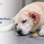
- Jaundice, visible yellowing of the eyes, gums, and ear skin, appearing when the thickened bile obstructs flow into the bile duct
- Increased thirst in dogs where an underlying endocrine condition is also present
- Fever in cases where infection or early rupture has occurred
The absence of dramatic early symptoms is what makes this condition dangerous. By the time severe signs appear, the gallbladder may already be critically compromised.
Why This Happens, The Underlying Triggers
Gallbladder mucocele does not develop in a vacuum. Several underlying conditions create the environment in which abnormal mucin production and bile stagnation occur.
Gallbladder dysmotility: The gallbladder depends on muscular contractions to empty bile into the bile duct. When these contractions are impaired or weakened, bile stagnates. Stagnant bile alters the chemical environment within the gallbladder, triggering mucus-secreting cells to overproduce mucin.
Endocrine disease: Hyperadrenocorticism (Cushing’s disease) and hypothyroidism are strongly associated with gallbladder mucocele development. Both conditions alter lipid metabolism, affect gallbladder motility, and create the internal conditions that favour mucin accumulation. Dogs with these diagnoses require regular biliary monitoring.
Hyperlipidaemia: Abnormally elevated blood lipid levels, whether due to diet, endocrine disease, or breed predisposition, are a recognised risk factor. High circulating lipids are thought to alter bile composition and gallbladder wall function.
Breed predisposition: Shetland Sheepdogs and Border Collies have a significantly elevated risk of developing mucocele. Cocker Spaniels and Miniature Schnauzers are also over-represented. Miniature Schnauzers, in particular, have a known predisposition to both hyperlipidaemia and biliary disease.
Biliary dysmotility with age: Older dogs show progressive decline in gallbladder motility in some cases, increasing the risk of bile stagnation and secondary mucocele formation.
Related Videos

How the Disease Develops Inside
The progression from early mucocele to life-threatening rupture follows a predictable biological sequence.
Stage 1: Bile stagnation Impaired gallbladder emptying causes bile to remain in the gallbladder longer than normal. As bile sits stagnant, its composition changes. Mucin-secreting goblet cells in the gallbladder wall respond to this altered environment by increasing mucus secretion.
Stage 2: Mucin accumulation Mucin mixes with the existing bile and begins to accumulate. The material thickens progressively. Bile that was once fluid becomes viscous, then gelatinous, then semi-solid.
Stage 3: Gallbladder expansion As the mucocele mass grows, the gallbladder expands under the increasing internal volume. The wall stretches. The characteristic stellate or kiwi-like pattern of the mucocele becomes visible on ultrasound at this stage.
Stage 4: Wall ischaemia Continued distension compresses the tiny blood vessels supplying the gallbladder wall. Areas of the wall begin to lose an adequate blood supply. Tissue becomes ischaemic and starts to lose its structural integrity.
Stage 5: Rupture risk Ischaemic wall tissue weakens to the point where it can no longer contain the pressure within. Rupture may occur suddenly and without any dramatic preceding escalation in symptoms. When the gallbladder ruptures, bile and mucocele contents spill into the abdominal cavity.
Stage 6: Bile peritonitis Bile in the abdominal cavity causes a severe, rapidly progressing inflammatory response. This is bile peritonitis. Without immediate surgical intervention, it is fatal.
Different Stages of the Condition, From Silent to Critical
Early stage: No clinical symptoms. The mucocele is present and visible on ultrasound, but the gallbladder has not yet reached a pressure point. Some early cases detected at this stage can be managed medically, though close monitoring is required.
Progressive stage: Clinical signs begin to appear as the mucocele expands. Vomiting, lethargy, and appetite loss reflect the increasing internal disruption. Liver enzymes begin to rise as the bile duct becomes partially obstructed or gallbladder inflammation affects adjacent liver tissue.
Advanced stage: The gallbladder is severely distended. Jaundice is visible from bile duct obstruction. The gallbladder wall shows signs of ischaemia on imaging. This is a pre-rupture state requiring surgical intervention without delay.
Ruptured stage: Acute, severe abdominal pain. Rapid deterioration. Fever. Collapse. Bile peritonitis in progress. This is a surgical emergency with a significantly higher mortality rate than elective surgery performed before rupture.
How Vets Confirm This Condition Accurately
Gallbladder mucocele has a distinctive appearance on ultrasound that allows experienced clinicians to identify it with high accuracy.
Ultrasound: The primary diagnostic tool. A healthy gallbladder appears as an anechoic (black) fluid-filled structure on ultrasound. A mucocele produces a characteristic stellate or kiwi-like pattern, with the gelatinous mucin showing as hyperechoic striated material within the gallbladder lumen. This appearance is considered pathognomonic for a mucocele. Ultrasound also assesses gallbladder wall thickness, integrity, and the presence of free abdominal fluid, suggesting rupture.
Blood panel: Liver enzyme elevation, including ALP and ALT, is common. ALP is frequently markedly elevated in mucocele cases, sometimes dramatically so. Elevated bilirubin confirms biliary obstruction. A complete blood count may show an elevated white cell count if infection or rupture has occurred.
Urinalysis: Bilirubinuria confirms hepatobiliary involvement. Urine colour may be abnormally dark.
Assessment of underlying endocrine disease: Given the strong association with Cushing’s disease and hypothyroidism, adrenal function testing and thyroid hormone measurement are indicated in all confirmed mucocele cases.
Treatment, Why Surgery Is Often the Only Real Solution
For most dogs with gallbladder mucocele, surgical removal of the gallbladder (cholecystectomy) is the definitive and recommended treatment.
Why surgery is the gold standard: Medical management cannot remove the gelatinous mucocele contents from the gallbladder. The substance does not dissolve with medication. The underlying dysmotility that created the condition persists. Without surgery, the mucocele continues to expand, and the rupture risk remains.
Pre-surgical stabilisation: Before surgery, dogs require intravenous fluid support to correct dehydration and electrolyte imbalances. Vitamin K is administered to assess and support clotting function, which is frequently compromised due to bile duct involvement and reduced fat-soluble vitamin absorption. Antibiotics are given as prophylaxis and to address any early infection.
Surgical procedure: Cholecystectomy involves the careful removal of the entire gallbladder, including all mucocele contents, with ligation of the cystic duct. The abdominal cavity is thoroughly lavaged. If bile peritonitis is present, abdominal drainage is also established. Dogs do not require a gallbladder to live normally. Bile flows directly from the liver through the hepatic ducts into the small intestine after gallbladder removal.
Medical management in early cases: In a small subset of early-stage cases with no wall compromise and minimal clinical signs, medical management with ursodiol to improve bile flow, dietary fat restriction, and treatment of the underlying endocrine disease may be considered with very frequent monitoring. This is not without risk and requires a clear owner-vet discussion about the ongoing rupture threat.
What Recovery Looks Like After Treatment
Dogs that undergo successful cholecystectomy before gallbladder rupture have a good long-term prognosis.
Post-operative care includes continued intravenous fluids, pain management, antibiotics, and gradual reintroduction of food over 24 to 48 hours. Liver enzyme levels are monitored to confirm they are trending downward. Most dogs show significant clinical improvement within 48 to 72 hours of successful surgery.
Long-term management includes:
- A low-fat diet to reduce lipid-related risk factors
- Treatment of any underlying endocrine disease
- Regular ultrasound monitoring of the biliary system, particularly in predisposed breeds
- Ongoing liver enzyme monitoring every three to six months
Dogs that undergo surgery after rupture have a significantly higher complication rate and a more guarded prognosis. The outcome depends on how quickly they reach surgery and how extensively bile peritonitis has progressed.
Complications That Make This Condition Dangerous
Gallbladder rupture: The most feared complication. May occur suddenly without dramatic preceding signs. Results in immediate bile peritonitis.
Bile peritonitis: Bile in the abdominal cavity causes chemical irritation and rapidly progressing bacterial peritonitis. Without surgical intervention within hours, this is fatal. Read more on this specific complication through VOSD’s guide on gallbladder obstruction in dogs for context on related biliary emergencies.
Bile duct obstruction: The expanding mucocele can compress or block the common bile duct, causing complete obstruction of bile flow, jaundice, and escalating liver damage.
Secondary hepatitis: Prolonged bile stasis and mucocele-driven inflammation damage adjacent liver tissue, causing secondary hepatitis and elevated liver enzymes.
Septic cholecystitis: Bacterial infection within the thickened mucocele contents causes suppurative cholecystitis, a severely inflamed, infected gallbladder that dramatically increases surgical risk.
Gallbladder Mucocele vs Gallstones vs Obstruction, Comparison
| Feature | Gallbladder Mucocele | Gallstones | Bile Duct Obstruction |
|---|---|---|---|
| Primary problem | Mucin accumulation in gallbladder | Mineral deposits in gallbladder | Mechanical blockage of bile duct |
| Cause | Dysmotility, endocrine disease | Abnormal bile composition | Stones, tumours, pancreatitis |
| Ultrasound appearance | Kiwi or stellate pattern | Hyperechoic foci with shadowing | Dilated bile duct |
| Rupture risk | High if untreated | Lower unless duct obstructed | Moderate |
| Treatment | Surgical removal usually required | Surgical or medical depending on size | Remove or bypass obstruction |
| Urgency | High, escalating to surgical emergency | Variable | High |
These three conditions can coexist. A mucocele can cause secondary obstruction. Gallstones can contribute to biliary stasis that worsens a mucocele. Accurate imaging and diagnosis are essential.
In dogs showing signs of systemic gastrointestinal disease alongside biliary findings, conditions such as inflammatory bowel disease in dogs may also need to be evaluated as concurrent or contributing factors.
When This Becomes an Immediate Surgical Emergency
Some presentations of gallbladder mucocele cannot wait for elective scheduling. They require emergency surgical intervention.
Go to the nearest veterinary clinic immediately if your dog:
- Collapses or is unable to stand
- Shows sudden severe abdominal pain, rigid abdomen, or screaming on touch
- Has visible jaundice alongside fever and rapid deterioration
- Was recently diagnosed with mucocele and has deteriorated acutely
- Has pale gums and a racing heart rate alongside abdominal signs
- Is vomiting continuously and completely unresponsive to supportive care
Suspected gallbladder rupture is a surgical emergency. Every hour of delay worsens the peritonitis and worsens the prognosis.
When You Should Stop Waiting and See a Vet Immediately
You do not need to reach a crisis level before acting. Seek veterinary attention promptly if:
- Your dog has been vomiting on and off for more than 24 hours with lethargy
- Is there any new yellowing visible in the eyes or gums?
- Your dog belongs to a predisposed breed and has not had recent abdominal imaging
- Your dog has a known diagnosis of Cushing’s disease or hypothyroidism and has not had a gallbladder assessment
- Appetite has been reduced for several days without a clear explanation
A mucocele detected before rupture is a manageable surgical condition. A mucocele discovered after rupture is an emergency with a significantly reduced margin for survival.