The immune system is supposed to protect the body.
And most of the time, it does exactly that. It identifies threats, surrounds them, and neutralises them. But sometimes, in its attempt to contain a problem it cannot fully resolve, it creates a different kind of damage.
That is precisely what happens in granulomatous hepatitis.
The body recognises something inside the liver as a persistent threat. It sends immune cells to deal with it. Those immune cells cluster together, forming small nodular structures called granulomas. And in doing so, they disrupt the liver tissue around them, impair normal function, and trigger a slow, complex inflammation that is notoriously difficult to diagnose and treat.
This is not a common condition. But when it occurs, it requires careful investigation because treating it incorrectly, or not treating it at all, leads to irreversible liver damage.
What This Condition Actually Is
Granulomatous hepatitis is a specific pattern of liver inflammation characterised by the formation of granulomas within liver tissue.
A granuloma is not a tumour. It is a tightly organised cluster of immune cells, primarily macrophages, that the immune system assembles around a substance or organism it cannot eliminate through its normal processes.
Think of it as the immune system building a wall around a problem it cannot solve. The wall contains the threat but does not remove it. And over time, multiple walls forming across the liver begin to interfere with how the organ functions.
Key points:
- Granulomas replace functional liver tissue wherever they form
- They trigger ongoing inflammation in the surrounding tissue
- They can be microscopic or large enough to be visible on imaging
- They are the liver’s response to a persistent, unresolved trigger
- The presence of granulomas in a liver biopsy is what distinguishes this condition from other forms of hepatitis
What You May Notice: Symptoms That Often Overlap With Other Diseases
One of the most challenging aspects of granulomatous hepatitis is that its symptoms are shared with many other conditions. There is no clinical sign that points uniquely to this diagnosis.
Common symptoms include:
- Intermittent or persistent vomiting
- Progressive weight loss without a dietary explanation
- Lethargy and reduced interest in normal activities
- Reduced or absent appetite
- Fever that comes and goes without an obvious source
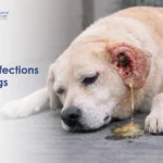
- Jaundice visible as yellowing of the eyes, gums, or skin
- Abdominal swelling from fluid accumulation in advanced cases
- Increased thirst and urination in some dogs
The combination of fever, weight loss, and vague digestive signs in a dog whose bloodwork shows elevated liver enzymes is a pattern that should prompt a deeper investigation rather than a presumptive diagnosis of more common liver conditions.
Why This Happens: The Underlying Triggers
Granuloma formation in the liver is always a response to something. Identifying what that something is determines the entire treatment approach.
Known causes include:
Fungal infections:
- The most common infectious cause of hepatic granulomas in dogs
- Histoplasma capsulatum, Blastomyces dermatitidis, and Cryptococcus neoformans are the most frequently identified organisms
- Fungal granulomas tend to be widespread and are associated with systemic illness
Bacterial infections:
- Mycobacterial infections, including atypical tuberculosis, can cause hepatic granulomas
- Brucellosis and certain other chronic bacterial infections are less common triggers
Parasitic causes:
- Larval migration of parasites through the liver can trigger granuloma formation
- Toxocara canis and other migrating larvae are occasional culprits
Cancer:
- Certain tumours, particularly lymphoma, can produce a granulomatous response in the liver
- This form is less responsive to treatment and carries a more guarded prognosis
Immune-mediated disease:
- The immune system attacks liver tissue without an identifiable infectious trigger
- This form, sometimes called idiopathic granulomatous hepatitis, is managed differently from infection-driven disease
Drug reactions:
- Certain medications can trigger granuloma formation as an adverse reaction
- Always inform your vet of every medication your dog is or has been receiving
Related Videos


Inside the Body: The Immune Reaction That Causes Damage
Understanding the mechanism explains why this condition is so difficult to resolve.
Here is the step-by-step progression:
- A persistent trigger, whether a fungal organism, bacterium, parasite, or unrecognised antigen, is detected inside liver tissue
- The immune system sends macrophages to engulf and destroy it
- When the macrophages cannot fully eliminate the trigger, they transform into larger cells called epithelioid cells and fuse
- These fused cells, surrounded by additional lymphocytes and other immune cells, form a compact nodule called a granuloma
- The granuloma persists as long as the trigger remains present
- Over time, multiple granulomas form across the liver, physically replacing functional hepatocytes
- The surrounding liver tissue becomes inflamed and progressively damaged
- If the process continues long enough, fibrosis develops around the granulomas, permanently scarring the affected areas
The body is not malfunctioning. It is doing exactly what it is designed to do. But the cumulative effect of that immune response inside a vital organ is organ damage.
When this granulomatous reaction triggers metabolic consequences, it can also affect the body’s overall acid-base balance. Dogs with significant liver dysfunction are at risk of developing complications, including lactic acid build-up, which adds another layer of clinical complexity to an already challenging condition.
Different Forms This Condition May Take
Granulomatous hepatitis is not a uniform diagnosis. It presents in different forms depending on the underlying cause.
Infectious granulomatous hepatitis:
- Driven by fungal, bacterial, or parasitic organisms
- Granulomas contain evidence of the causative organism on special staining
- Treatment is directed at the underlying infection
- Prognosis is better if the infection is treatable and caught early
Non-infectious or immune-mediated granulomatous hepatitis:
- No organism is identified within the granulomas
- The immune system appears to be reacting to the liver’s own tissue or to an unidentified antigen
- Requires immunosuppressive therapy
- Can be more difficult to control long-term
Idiopathic granulomatous hepatitis:
- Granulomas are present, but no cause is found despite thorough investigation
- Managed empirically based on clinical response to treatment
- Regular monitoring is essential
Pyogranulomatous hepatitis:
- A mixed pattern where both granulomas and suppurative inflammation are present simultaneously
- Often associated with certain bacterial infections
- Requires a specific treatment approach targeting both components
Why Diagnosis Is Often Difficult and Delayed
Granulomatous hepatitis is one of the more diagnostically challenging liver conditions in dogs.
The reasons for diagnostic difficulty include:
- The early symptoms are non-specific and shared with many common conditions
- Elevated liver enzymes on bloodwork alone do not distinguish this from other hepatitis patterns
- Imaging can show liver abnormalities, but cannot identify granulomas definitively
- A definitive diagnosis requires liver biopsy with histopathology and special staining
- Even after a biopsy confirms granulomas, identifying the underlying cause requires additional testing, including fungal cultures, PCR, serology, and, in some cases, bone marrow evaluation
Diagnostic steps your vet will typically follow:
- Full blood panel including liver enzymes, bile acids, albumin, and clotting times
- Abdominal ultrasound to assess liver architecture and identify any masses or enlarged lymph nodes
- Chest radiographs if fungal infection is suspected, as many fungal diseases also affect the lungs
- Liver biopsy under ultrasound guidance or during exploratory surgery
- Special tissue stains to identify fungal organisms, acid-fast bacteria, or parasitic material
- Systemic fungal antigen testing where available
Without a biopsy, a presumptive diagnosis is often made based on the clinical picture and the dog’s response to empirical treatment.
Treatment Depends on Finding the Root Cause
This is the most important principle in managing granulomatous hepatitis. Treating the inflammation without addressing the underlying trigger rarely leads to lasting improvement.
For fungal causes:
- Long-term antifungal therapy with itraconazole, fluconazole, or amphotericin B, depending on the organism and disease severity
- Treatment duration is typically several months and guided by antigen testing and clinical response
For bacterial causes:
- Prolonged antibiotic therapy targeted to the specific organism identified on culture
- Mycobacterial infections require combination antibiotic protocols
For immune-mediated or idiopathic forms:
- Immunosuppressive therapy with prednisolone, often at higher doses initially
- Azathioprine may be added in cases requiring stronger immune suppression
- Treatment is tapered slowly based on clinical and laboratory response
Supportive care for all forms:
- Liver-supportive supplements, including SAMe and milk thistle, to reduce oxidative stress
- Ursodeoxycholic acid to improve bile flow
- Diet modification to reduce liver workload
- Fluid therapy and hospitalisation in dogs with significant deterioration
Regular monitoring through repeat bloodwork and imaging is essential throughout treatment to assess response and catch deterioration early.
What Recovery Looks Like, And Why It Varies So Much
Prognosis for granulomatous hepatitis depends almost entirely on two factors: the underlying cause and how early treatment begins.
Better prognosis scenarios:
- Dogs with infectious granulomatous hepatitis caused by a treatable organism caught before significant fibrosis
- Dogs with immune-mediated disease that responds to immunosuppressive therapy
- Cases where treatment is started before the granulomatous process has spread extensively
More guarded prognosis scenarios:
- Dogs presenting with advanced fibrosis or early cirrhosis at the time of diagnosis
- Cases where no underlying cause is identified and response to empirical treatment is poor
- Cancer-associated granulomatous hepatitis, particularly lymphoma-driven cases
Young dogs diagnosed with liver complications face particular challenges. The guide on liver fibrosis in young dogs explains how early scarring processes develop and why age at diagnosis affects long-term outcomes.
What Happens If the Underlying Cause Is Not Treated
Without addressing the root trigger, granulomatous hepatitis progresses in a predictable and damaging direction:
- Granulomas continue forming, progressively replacing functional liver tissue
- Fibrosis develops around existing granulomas and spreads through the liver
- Portal hypertension develops as scarring distorts normal blood flow
- Cirrhosis becomes established, at which point liver damage is irreversible
- Hepatic encephalopathy develops as the liver loses its ability to filter ammonia
- Fluid accumulates in the abdomen
- Liver failure and death follow without intervention
The immune response that formed the granulomas does not switch off on its own. It continues as long as the trigger is present. And with every granuloma that forms, more liver tissue is lost permanently.
Granulomatous Hepatitis vs Chronic Hepatitis vs Liver Tumours
| Feature | Granulomatous Hepatitis | Chronic Hepatitis | Liver Tumours |
|---|---|---|---|
| Primary mechanism | Immune granuloma formation | Persistent inflammatory cell infiltration | Abnormal cell proliferation |
| Common causes | Fungal, bacterial, immune, cancer | Immune, copper, toxins, infection | Primary or metastatic cancer |
| Biopsy appearance | Distinct granuloma nodules | Diffuse inflammatory infiltrate | Neoplastic cells |
| Reversibility | Partial if cause is treated early | Partial if caught before cirrhosis | Depends on tumour type and stage |
| Treatment approach | Cause-specific plus supportive | Anti-inflammatory plus cause-specific | Surgical, chemotherapy, palliative |
| Neurological signs | In advanced cases | In advanced cases | Can occur with metastatic disease |
When This Condition Becomes an Emergency
Go to a veterinary clinic immediately without delay if your dog shows:
- Sudden onset of seizures or disorientation
- Rapid abdominal distension developing over hours
- Visible jaundice appearing quickly
- Collapse or inability to stand
- Complete refusal to eat or drink for more than 24 hours alongside fever or neurological signs
These signs indicate the liver has lost critical functional capacity and the dog requires emergency hospitalisation and stabilisation.
When You Should Not Wait
Even without emergency signs, schedule a veterinary appointment within 24 to 48 hours if your dog has:
- Fever without an obvious source that has persisted for more than a few days
- Unexplained weight loss combined with reduced appetite
- Vomiting that keeps returning despite treatment for simpler causes
- Elevated liver enzymes found on routine bloodwork without a clear explanation
- Any combination of digestive signs and lethargy that is not improving
For a broader reference on the full range of medical conditions that can cause similar clinical pictures, the VOSD dog medical conditions library provides comprehensive coverage of conditions your vet may consider in the differential diagnosis.