Most liver conditions in dogs develop slowly. They build over months, sometimes years, before symptoms become obvious.
Suppurative hepatitis does not follow that pattern.
This is an infection-driven condition where bacteria invade liver tissue, and the body’s immune response creates pus-filled pockets called abscesses within the organ itself. It moves quickly. It escalates without warning. And by the time the signs are obvious enough to bring a dog to the vet, the infection may already be spreading beyond the liver.
This is not a condition to observe and wait on. It is one to act on the moment it is suspected.
What This Condition Really Is, Beyond Inflammation
Suppurative hepatitis is a bacterial infection of the liver that results in the formation of pus within liver tissue.
The word suppurative means pus-forming. Unlike inflammatory liver conditions driven by immune-mediated processes or chronic toxin exposure, suppurative hepatitis has a specific infectious origin. Bacteria invade the liver, the immune system responds by sending large numbers of neutrophils to the site of infection, and as those white blood cells fight the bacteria, tissue is destroyed, and pus accumulates.
When the infection is localised, discrete pockets of pus form within the liver tissue. These are called hepatic abscesses. When the infection is more diffuse, it can spread through the biliary system and liver tissue more broadly, a pattern that overlaps with infectious cholangiohepatitis.
In either case, the liver is not simply inflamed. It is being actively destroyed from within by an infectious process.
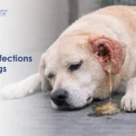
What You May Notice, Signs That Often Appear Suddenly
Unlike many liver conditions that creep up gradually, suppurative hepatitis frequently causes a sudden and significant change in a dog’s condition.
Signs commonly seen:
- Fever, often high and persistent, is one of the most consistent indicators of an active infection
- Marked lethargy, far more pronounced than typical tiredness
- Vomiting, which may begin suddenly and become frequent
- Complete or near-complete loss of appetite
- Abdominal pain, visible as hunching, reluctance to move, guarding the belly, or vocalising when touched in the abdominal region
- Jaundice, yellowing of the eyes, gums, and inner ear skin, when infection has disrupted bile flow
- Dehydration from vomiting, reduced drinking, and fever-driven fluid loss
- Rapid deterioration over 24 to 48 hours in some cases
The combination of fever and acute abdominal pain in any dog is a signal that demands same-day veterinary attention.
Why This Happens, How Infection Reaches the Liver
The liver is not directly exposed to the external environment. Bacteria reach it through several internal pathways.
Ascending biliary infection: Bacteria from the intestine travel upward through the bile duct into the biliary system and from there into the liver. This is the most common route. It explains why dogs with pre-existing bile duct disease or chronic gastrointestinal conditions carry an elevated risk.
Haematogenous spread: Bacteria circulating in the bloodstream from an infection elsewhere in the body, a wound, a dental abscess, a gut infection, can be filtered by the liver and seed within hepatic tissue. The liver processes all blood from the portal circulation, making it one of the most exposed organs to blood-borne pathogens.
Direct trauma: Penetrating wounds or surgical complications can introduce bacteria directly into liver tissue or its surroundings.
Tumour-associated infection: Necrotic tissue within hepatic tumours creates an environment where bacteria thrive. Secondary infection within a tumour mass can progress into suppurative hepatitis.
Immune compromise: Dogs on long-term immunosuppressive medications, dogs with systemic disease affecting immune function, or dogs in poor nutritional condition have reduced capacity to prevent opportunistic bacterial infection within the liver.
Related Videos


Inside the Body, The Infection Cascade That Damages the Liver
This section explains the mechanism that makes suppurative hepatitis so destructive when left untreated.
Step 1: Bacterial invasion Bacteria enter liver tissue through one of the pathways described above. Common bacteria involved include E. coli, Clostridium species, Streptococcus, Staphylococcus, and anaerobic organisms.
Step 2: Immune activation The liver’s immune cells, particularly Kupffer cells, recognise the bacterial invasion and trigger an acute inflammatory response. Signals are sent to recruit neutrophils, the primary white blood cells of acute infection response.
Step 3: Neutrophil accumulation Large numbers of neutrophils flood the affected area of the liver. Their job is to engulf and destroy bacteria.
Step 4: Pus formation As neutrophils fight the bacteria, both the bacteria and the white blood cells die and break down. This cellular debris, mixed with inflammatory fluid and dead tissue, forms pus. If the infection is localised, this pus becomes walled off by surrounding tissue into a discrete abscess.
Step 5: Tissue destruction The inflammatory process and direct bacterial toxins destroy the hepatocytes in the affected zone. Liver tissue is replaced by infected, non-functional material. If the abscess expands or multiple abscesses form, functional liver capacity declines.
Step 6: Systemic spillover If the infection is not controlled, bacteria and their toxins enter the bloodstream from the infected liver tissue, causing bacteraemia or sepsis. The systemic inflammatory response triggers fever, cardiovascular instability, and multi-organ involvement.
Different Forms This Condition May Present As
Suppurative hepatitis does not always look the same clinically or anatomically.
Localised hepatic abscess: A discrete, walled-off pocket of infection within one part of the liver. May be solitary or multiple. On ultrasound, it appears as a fluid-filled or mixed-echogenicity lesion. May be amenable to surgical drainage or aspiration alongside antibiotic therapy.
Diffuse suppurative hepatitis: Infection spreads more broadly through hepatic tissue and the biliary system. No discrete abscess is present, but neutrophilic infiltration is extensive throughout the liver. This form carries a more serious prognosis because there is no single surgical target, and the overall organ is compromised.
Suppurative cholangiohepatitis: Where infection has originated in or spread through the bile duct system into the liver. The biliary tree shows inflammation alongside hepatic infection. This is the form most commonly associated with ascending gut bacteria.
How Veterinarians Identify the Infection Source
Accurate diagnosis requires more than confirming that the liver is inflamed. The infectious nature of the inflammation must be established, and the source identified to guide treatment.
Blood panel: Marked elevation of liver enzymes, including ALT and ALP. Elevated white blood cell count, particularly neutrophils, strongly suggests active bacterial infection. Elevated bilirubin if biliary involvement is present. Hypoalbuminaemia in severe cases. Blood culture if bacteraemia is suspected.
Urinalysis: Assesses kidney involvement and biliary spillover. Bilirubinuria and abnormal urine colour may be present.
Abdominal ultrasound: The primary imaging tool. Hepatic abscesses appear as fluid-filled, mixed-echogenicity, or cavitated lesions within the liver parenchyma. Ultrasound also assesses bile duct involvement, free abdominal fluid, and its impact.
Ultrasound-guided fine needle aspiration: Collecting fluid directly from a suspected abscess for cytology and bacterial culture. This is both diagnostic and, in some cases, partially therapeutic if the pus volume is reduced. Culture results guide specific antibiotic selection.
Exploratory surgery or laparoscopy: In cases where imaging is inconclusive or where surgical drainage is required, direct visualisation and sampling of the affected liver tissue provides a definitive diagnosis.
Treatment Requires Immediate Infection Control
Suppurative hepatitis demands aggressive treatment. Supportive care alone is insufficient.
Intravenous antibiotics: The cornerstone of treatment. Broad-spectrum intravenous antibiotics are started immediately, before culture results are available, to cover the most likely bacterial organisms. Once culture and sensitivity results return, the antibiotic protocol is refined to target the specific bacteria identified. Treatment duration is typically a minimum of six to eight weeks, and often longer.
Intravenous fluid therapy: Corrects dehydration, supports blood pressure, and assists in flushing bacterial toxins from the circulation. Dogs with fever and vomiting are frequently significantly dehydrated at presentation.
Abscess drainage: Where a discrete abscess is identified, drainage reduces the bacterial load and pus volume within the liver. This may be achieved through ultrasound-guided aspiration or through surgical drainage, depending on abscess size, location, and accessibility.
Surgical intervention: In cases with large, solitary abscesses, multiple abscesses, or abscesses not responding to medical management, partial liver lobectomy, removing the affected portion of the liver, may be necessary. The liver has significant regenerative capacity and can tolerate the removal of a portion of its tissue.
Nutritional support: Dogs with suppurative hepatitis are often anorexic and catabolic. Early nutritional support through a feeding tube or parenteral nutrition maintains the metabolic resources the liver needs for recovery and immune function.
Management of the underlying source: Where the source of infection is identified, such as a biliary stricture, intestinal infection, or distant abscess, treating that source is essential to prevent recurrence.
What Recovery Depends On, And What to Expect
Prognosis is directly linked to how early treatment begins and whether the infection is controlled before systemic spread occurs.
Dogs treated promptly with appropriate antibiotics and supportive care, particularly those with a solitary, accessible abscess, can recover well. Liver enzyme levels normalise over weeks to months as the infection resolves and the liver regenerates.
Dogs in whom diagnosis is delayed, where the infection has spread to the bloodstream, or where multiple abscesses have destroyed a significant portion of liver tissue, face a more guarded prognosis. Sepsis significantly worsens outcomes.
Long-term monitoring with regular blood panels and ultrasound is required to confirm resolution and detect early recurrence.
What Happens If the Infection Spreads or Is Untreated
The consequences of delayed or absent treatment escalate rapidly.
- Sepsis: Bacteria entering the bloodstream from the infected liver trigger a systemic inflammatory response. Septic shock involves cardiovascular collapse, organ failure, and carries a high mortality rate without intensive care.
- Liver failure: Extensive tissue destruction from multiple abscesses or diffuse suppurative infection eliminates functional liver tissue beyond the organ’s regenerative capacity.
- Abscess rupture: A large hepatic abscess can rupture into the abdominal cavity, causing septic peritonitis, a rapidly fatal complication.
- Multi-organ failure: Sustained sepsis and hepatic failure together stress the kidneys, cardiovascular system, and coagulation pathways simultaneously.
Suppurative Hepatitis vs Other Liver Inflammations
| Feature | Suppurative Hepatitis | Granulomatous Hepatitis | Chronic Hepatitis |
|---|---|---|---|
| Primary driver | Bacterial infection | Immune or fungal response | Immune-mediated, toxic, or genetic |
| Inflammatory cell | Neutrophils (pus-forming) | Macrophages (granuloma formation) | Lymphocytes and plasma cells |
| Onset | Acute, often sudden | Subacute to chronic | Insidious, progressive |
| Fever | Prominent | Variable | Uncommon |
| Abscess formation | Yes | No | No |
| Treatment | Antibiotics, drainage | Anti-fungal or immunosuppressive | Immunosuppressive, supportive |
| Urgency | High | Moderate | Moderate |
Getting this distinction right determines whether a dog receives antibiotics or immunosuppressing drugs. Giving immunosuppressives to a dog with an active bacterial infection is dangerous.
When This Condition Becomes an Emergency
Some presentations of suppurative hepatitis are not situations for a scheduled appointment.
Go to the nearest veterinary clinic immediately if your dog:
- Has a high fever alongside abdominal pain and vomiting
- Has collapsed or cannot stand
- Shows rapid deterioration over a few hours
- Has severe jaundice combined with fever and lethargy
- Is showing neurological signs, including disorientation or seizures
- Was recently diagnosed with a liver condition and has deteriorated acutely
High fever with abdominal pain in any dog is an emergency. It should never be observed overnight at home.
How This Condition Connects to Other Disorders
Suppurative hepatitis rarely exists in isolation. It is frequently connected to other hepatic and systemic conditions.
Understanding related conditions, including liver fistula in dogs, provides important context for how abnormal vascular and structural liver pathology can create conditions where infection takes hold. A broader view of dog medical conditions helps owners understand how interconnected organ systems are and why a liver infection can trigger problems far beyond the liver itself. For comprehensive guidance across all aspects of canine health, VOSD maintains an extensive resource library built around clinical accuracy and owner education.
When You Should Seek Immediate Veterinary Care
Do not wait if your dog shows:
- Any combination of fever, vomiting, and abdominal discomfort lasting more than a few hours
- Sudden and severe reduction in energy with no clear cause
- Any yellowing of the eyes or gums alongside the above signs
- Rapid deterioration after being mildly unwell the day before
Suppurative hepatitis is not a condition where a wait-and-see approach is safe. The window between early treatment and critical complications is narrow.