

We often see dogs brought in after a sudden collapse that resolved on its own within seconds. The dog is alert again, perhaps a little wobbly, and the owner is shaken and unsure what happened. In some cases, it happens once and is never seen again. In others, it becomes a pattern that points toward something happening in the heart.
Ventricular tachycardia in dogs is one of the more important cardiac arrhythmias to understand, because it sits on a wide spectrum. At one end, occasional brief episodes in a dog with otherwise mild heart disease may be monitored without immediate treatment. At the other end, sustained ventricular tachycardia is a life-threatening emergency requiring urgent intervention.
Knowing what ventricular tachycardia in dogs looks like, which dogs are at risk, and when to act gives you the foundation to respond well rather than be caught off guard.
What Is Ventricular Tachycardia in Dogs?
Ventricular tachycardia in dogs is a heart rhythm disorder in which abnormal electrical signals originating in the ventricles, the lower pumping chambers of the heart, cause the heart to beat far faster than it should. A normal resting heart rate for a dog is typically between 60 and 140 beats per minute depending on size and breed. During ventricular tachycardia, the rate can exceed 180 to 200 beats per minute or higher.
At such a rapid rate, the ventricles do not have enough time to fill properly between beats. Less blood is pumped with each contraction, and the body begins to receive inadequate circulation. This is what causes the clinical signs that pet parents and vets observe.
Ventricular tachycardia can be intermittent, occurring in short runs of rapid beats before returning to normal, or it can be sustained, continuing for minutes or longer. Sustained ventricular tachycardia is significantly more serious and requires immediate veterinary attention.
Causes of Ventricular Tachycardia in Dogs
Ventricular tachycardia in dogs rarely develops in isolation. It is almost always triggered by an underlying condition that has disrupted the electrical stability of the heart muscle. Identifying that underlying cause is central to managing the condition effectively.
| Cause Category | Examples |
|---|---|
| Structural heart disease | Dilated cardiomyopathy, valvular disease, myocarditis |
| Systemic and infectious illness | Tick-borne diseases, sepsis, pancreatitis, splenic disease |
| Electrolyte imbalances | Low potassium, low magnesium, high potassium from kidney failure |
| Trauma | Blunt chest trauma, post-surgical cardiac stress |
| Toxin exposure | Certain plants, drugs, anaesthetic agents, household chemicals |
| Tumours | Cardiac tumours, splenic haemangiosarcoma with cardiac involvement |
| Idiopathic | No identifiable cause; occasionally seen in young large breed dogs |
In the Indian context, tick-borne diseases deserve particular mention. Ehrlichiosis, babesiosis, and related infections cause systemic illness that can destabilise cardiac rhythm, particularly when they go unrecognised or undertreated for a period. A dog with a history of tick exposure who develops fainting or collapse should have tick-borne disease screening as part of the initial work-up, alongside cardiac evaluation.
Blunt trauma is another trigger that is sometimes overlooked. A dog struck by a vehicle, even if they appear relatively uninjured externally, can develop ventricular arrhythmias in the 24 to 72 hours following the incident. Any dog involved in a road accident should be monitored with an ECG for at least 24 hours regardless of their outward appearance.
Related Products


Common Risk Factors
- Large and giant breed dogs, particularly Dobermann Pinschers, Boxers, and German Shepherds, which have higher rates of dilated cardiomyopathy
- Dogs with known structural heart disease or previous myocarditis
- Dogs with chronic kidney disease causing electrolyte disturbances
- Older dogs with multiple concurrent health conditions
- Dogs with significant tick exposure and inadequate parasite prevention
- Dogs who have experienced recent trauma or major surgery
Symptoms of Ventricular Tachycardia in Dogs
The symptoms of ventricular tachycardia in dogs depend on how fast the heart is beating, how long episodes last, and how well the rest of the cardiovascular system is compensating. Brief, self-terminating runs may produce minimal or no noticeable signs. Sustained or recurrent episodes cause more obvious and concerning effects.
| Episode Type | Likely Symptoms |
|---|---|
| Brief, intermittent runs | Momentary weakness, brief wobbliness, mild restlessness, occasional pause in activity |
| Frequent or longer runs | Exercise intolerance, lethargy, reduced appetite, panting at rest, apparent anxiety |
| Sustained tachycardia | Fainting or syncope, collapse, pale or grey gums, extreme weakness, rapid shallow breathing |
| Severe or degenerating | Complete collapse, unresponsiveness, absent or barely perceptible pulse |
Episodes can be brief but important to notice and track. A dog that momentarily stumbles, sits down suddenly, appears glassy-eyed for a few seconds, and then seems fine may be experiencing a brief run of ventricular tachycardia. These episodes are easily dismissed, particularly if they do not recur immediately, but a pattern of such events in a dog with any of the known risk factors should be investigated.
In some cases, fainting associated with ventricular tachycardia can look similar to a seizure, with brief muscle twitching or rigidity as the dog loses consciousness. The distinction matters because the management is very different. A vet assessment including an ECG is the way to determine what is actually happening.
When Is It an Emergency?
The following signs require immediate veterinary attention. Do not wait for a scheduled appointment:
- Collapse that lasts more than a few seconds or is followed by obvious weakness
- Repeated fainting or collapse episodes within a short period
- Pale, grey, or bluish gums at any point
- Rapid, shallow, or laboured breathing at rest
- Complete unresponsiveness, even briefly
- Rapid heart rate you can feel on the chest that does not slow down
Keep your dog as still and calm as possible, avoid stimulating them, and get to a vet immediately. Call ahead if you can so the team is ready on arrival.
Related Videos


Diagnosis of Ventricular Tachycardia in Dogs
Diagnosing ventricular tachycardia in dogs requires cardiac testing. Clinical signs alone are not specific enough to confirm the arrhythmia or distinguish it from other causes of collapse or weakness.
The main diagnostic tools include:
- Electrocardiogram (ECG): This is the definitive diagnostic test. An ECG records the heart’s electrical activity and can identify the rapid, wide, abnormal complexes characteristic of ventricular tachycardia. If an episode is occurring at the time of recording, diagnosis is straightforward. If episodes are intermittent, the ECG may be normal between events.
- Holter monitor: A portable ECG worn at home for 24 to 48 hours. This is particularly valuable for catching intermittent episodes that do not appear during a brief clinic visit. The recording is reviewed afterwards to identify abnormal rhythms and assess their frequency and duration.
- Echocardiography: Cardiac ultrasound evaluates the structure and function of the heart, identifies any underlying cardiomyopathy or other structural disease, and assesses the degree of cardiac compromise.
- Blood tests: A full biochemistry panel checks electrolytes, kidney and liver function, and overall metabolic status. Tick-borne disease panels are important in India given the prevalence of tick exposure.
- Chest radiographs: To assess heart size and lung changes consistent with early heart failure.
Access to Holter monitoring and echocardiography varies across India. Larger cities generally have specialist veterinary cardiac services. In areas with more limited access, a combination of ECG, blood work, and careful clinical assessment provides a useful starting point while referral is arranged if needed.
Treatment for Ventricular Tachycardia in Dogs
Treatment for ventricular tachycardia in dogs has two priorities: stabilising the heart rhythm when it is causing clinical signs, and addressing the underlying condition responsible for triggering the arrhythmia.
Emergency Stabilisation
When a dog arrives in active, sustained ventricular tachycardia, the immediate goal is to slow and stabilise the heart rate. This typically involves:
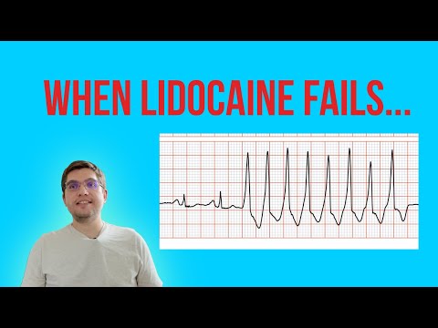
- Intravenous anti-arrhythmic medication, most commonly lidocaine as a bolus and then as a continuous rate infusion, to interrupt the abnormal rhythm
- Continuous ECG monitoring to assess response to treatment in real time
- Supplemental oxygen to support circulation
- Intravenous fluids if the dog is haemodynamically compromised
- Correction of any electrolyte abnormalities identified on blood work
Most dogs respond well to intravenous lidocaine when ventricular tachycardia is the primary issue. If the rhythm does not convert, other agents may be added. The goal is to convert to a normal sinus rhythm and then maintain stability while the underlying cause is investigated.
Long-Term Medical Management
Once stabilised, many dogs with ventricular tachycardia require ongoing oral anti-arrhythmic medication to prevent recurrence. Commonly used agents include mexiletine, sotalol, and atenolol, often in combination depending on the specific arrhythmia pattern and the dog’s overall cardiac status.
Treating the underlying cause is equally important:
| Underlying Cause | Primary Treatment Focus |
|---|---|
| Dilated cardiomyopathy | Cardiac medications, anti-arrhythmics, regular monitoring |
| Tick-borne disease | Doxycycline course, supportive care, cardiac monitoring |
| Electrolyte imbalance | Correction of potassium or magnesium, treat underlying cause |
| Post-trauma arrhythmia | Hospitalisation, monitoring, anti-arrhythmics as needed |
| Splenic or cardiac tumour | Surgical assessment, palliative management depending on case |
| Toxin exposure | Decontamination, supportive care, cardiac monitoring |
Home Care and Monitoring
For dogs managed on long-term anti-arrhythmic medication, consistent home care makes a meaningful difference:
- Administer medication at the same time each day without missing doses. Anti-arrhythmic medication loses effectiveness quickly if doses are skipped.
- Keep a simple log of any episodes of weakness, stumbling, or collapse, including the time, duration, and what the dog was doing beforehand.
- Monitor resting breathing rate as a general indicator of cardiac stability.
- Restrict vigorous exercise until your vet confirms it is safe to gradually increase activity.
- Keep the home environment calm, particularly during periods of adjustment to new medication.
Related Products


Prognosis: What to Expect
The prognosis for ventricular tachycardia in dogs is variable and depends substantially on the underlying cause, the frequency and severity of episodes, and how quickly treatment is started.
Dogs whose ventricular tachycardia is caused by a reversible or treatable condition, such as an electrolyte imbalance, tick-borne disease, or post-traumatic arrhythmia, often have a very good outcome once the underlying problem is addressed. The arrhythmia may resolve entirely once the root cause is treated.
Dogs with ventricular tachycardia caused by dilated cardiomyopathy or other progressive cardiac disease face a more guarded long-term outlook, though appropriate anti-arrhythmic treatment significantly improves quality of life and may extend survival time. Regular follow-up ECGs and echocardiograms allow treatment to be adjusted as the condition evolves.
The most important factor across all cases is timely recognition and consistent management. Dogs whose arrhythmia is caught early and managed carefully consistently do better than those where treatment is delayed or inconsistent.
Preventing Ventricular Tachycardia in Dogs
Ventricular tachycardia cannot always be prevented, particularly when it arises from a genetic cardiac predisposition. However, several measures reduce the risk in susceptible dogs:
- Consistent tick and flea prevention to reduce the risk of tick-borne disease reaching the heart
- Prompt veterinary treatment of any systemic infection or illness before it progresses
- Annual health checks including cardiac auscultation for dogs aged seven and above, or from a younger age in predisposed breeds
- Safe storage of all medications, chemicals, and potentially toxic plants away from dogs
- ECG monitoring for at least 24 hours after any significant blunt trauma, even in a dog that appears stable
Living with a Dog with a Heart Rhythm Disorder
Managing a dog with ventricular tachycardia long-term asks consistency and attentiveness from the whole household. It does not need to be complicated, but it does need to be steady.
Most dogs on well-managed anti-arrhythmic treatment settle into a comfortable routine. Their activity is adjusted rather than eliminated. Their medication becomes part of the daily rhythm of the house. Their owners become skilled at recognising what a good day looks like versus a day that needs a call to the vet.
The families that achieve the best outcomes are those who stay in close contact with their vet, report changes promptly rather than waiting to see if things improve, and approach the condition with calm consistency rather than anxiety. Your steadiness genuinely transfers to your dog.
Ventricular tachycardia in dogs is a condition that rewards early recognition, calm assessment, and consistent management. It is not a single event to be fixed and forgotten. It is a pattern to be understood, monitored, and managed over time with the right support.
Whether your dog has experienced a single unexplained fainting episode or has already been diagnosed with ventricular tachycardia in dogs, the path forward is the same: work closely with your vet, stay consistent with medication and monitoring, and observe your dog with the kind of steady attentiveness that makes a real difference. Early action and consistent care change outcomes in ways that matter.


