

A puppy can be born with a serious eye condition and show no visible sign of it whatsoever.
No redness. No discharge. No squinting. Nothing that would make even an attentive owner pause. And yet, behind those apparently normal eyes, the structures responsible for vision may already be malformed, underdeveloped, or damaged in ways that cannot be reversed.
This is the nature of Collie Eye Anomaly. It is inherited, congenital, and in many cases, completely silent until a veterinary eye examination reveals what the naked eye never could.
CEA is not an infection. It is not caused by an injury or an environmental trigger. It is written into the dog’s genetics before birth, which is precisely what makes it one of the most important conditions for owners and breeders of affected breeds to understand.
What is Collie Eye Anomaly?
Collie Eye Anomaly, commonly referred to as CEA, is a hereditary developmental eye disease that affects the formation of the eye during a puppy’s development in the womb. It is present from birth and affects the choroid, the layer of blood vessels beneath the retina that supplies oxygen and nutrients to the eye tissue.
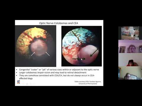
In CEA, the choroid does not develop properly. This is called choroidal hypoplasia, and it is the hallmark finding of the condition. Depending on severity, the disease may also involve the retina, the optic disc, and the sclera, the white outer layer of the eye.
One of the most important clinical features of CEA is that it affects both eyes in virtually all cases, but not necessarily to the same degree. One eye may have a mild form while the other is more severely affected. This asymmetry can make the condition easy to miss without a thorough ophthalmic examination.
CEA is not an inflammatory condition, and it is not contagious. It is a structural developmental failure, and no amount of medication, eye drops, or dietary intervention will change what has already formed in the tissue.
Symptoms of Collie Eye Anomaly
The range of symptoms in CEA is wide, and that range is part of what makes this condition clinically deceptive.
Mild Cases
Many dogs with mild CEA show no symptoms at all. They navigate their environment normally, respond to visual cues, and appear completely unaffected in daily life. The only abnormality is visible on ophthalmoscopic examination, where the choroidal hypoplasia can be seen by a veterinary ophthalmologist as a pale, poorly pigmented area beneath the retina.
These dogs are often described as having “go normal” CEA, a term used when the disease is so mild that areas of choroidal hypoplasia become pigmented over time and harder to detect. This does not mean the condition has resolved. It means it has become masked, which is why early screening at six to eight weeks of age is critical.
Visual Deficits
Dogs with moderate CEA may show behavioural signs of reduced vision that owners sometimes attribute to clumsiness or personality.
- Hesitation at steps, thresholds, or unfamiliar environments
- Bumping into objects, particularly in low light conditions
- Reluctance to navigate new spaces
- Difficulty tracking moving objects
- Reduced responsiveness to visual cues at a distance
If a dog that has always seemed slightly cautious or visually uncertain comes from a breed predisposed to CEA, an eye examination is warranted.
Structural Signs
In more pronounced cases, changes in the physical appearance of the eye may be visible.
- Microphthalmia, where the eye is noticeably smaller than normal
- A sunken or deep-set appearance to one or both eyes
- Cloudiness or opacity in the eye
- Abnormal position or appearance of the pupil
These structural signs indicate more significant developmental disruption and are associated with more severe visual impairment.
Severe Signs
In the most serious cases of CEA, retinal detachment occurs. This is when the retina separates from the underlying tissue, cutting off its blood supply and causing sudden and often permanent vision loss. Retinal detachment can affect one or both eyes and may occur without warning.
Dogs with retinal detachment may suddenly begin walking into objects they previously navigated without difficulty, appear disoriented, or show a visible change in the appearance of the affected eye.
Sudden vision loss in any dog is a veterinary emergency.
Causes of Collie Eye Anomaly
Genetic Mutation
CEA is caused by a mutation in the NHEJ1 gene, inherited in an autosomal recessive pattern. For a dog to be affected by CEA, it must inherit a copy of the mutated gene from both parents. A dog that inherits the mutation from only one parent is a carrier. Carriers do not develop CEA themselves but can pass the gene on to their offspring.
This inheritance pattern means that two apparently healthy, visually normal dogs can produce affected puppies if both are carriers of the mutation. Without genetic testing, there is no way to know whether a breeding dog is a carrier.
Developmental Failure in the Womb
The structural abnormalities of CEA arise during a specific window of eye development in the embryo. The choroid fails to develop its full complement of blood vessels and tissue, and this failure is permanent. By the time the puppy is born, the developmental window has already closed, and the eye structure is already fixed.
Breed Predisposition
CEA has the highest prevalence in herding breeds. The breeds most commonly affected include:
- Rough and Smooth Collies
- Border Collies
- Shetland Sheepdogs (Shelties)
- Australian Shepherds
- Lancashire Heelers
- Nova Scotia Duck Tolling Retrievers
In some collie populations, studies have found CEA affecting a significant proportion of dogs. This makes genetic screening not just recommended but essential in these breeds, both for individual health decisions and responsible breeding programmes.
Related Videos


Diagnosis of Collie Eye Anomaly
Early diagnosis is one of the most important aspects of managing CEA, both for the individual dog and for responsible breeding decisions.
Ophthalmic Examination at Six to Eight Weeks
The ideal window for diagnosing CEA through ophthalmoscopy is between six and eight weeks of age. During this period, the characteristic pale areas of choroidal hypoplasia are most visible before natural pigmentation begins to obscure them. A veterinary ophthalmologist uses an instrument called an indirect ophthalmoscope to examine the retina and choroid directly.
After this window, mild cases may become harder to detect through examination alone, which is why early screening is not optional in at-risk breeds. For a broader context on inherited structural conditions affecting the eye, our guide on congenital eye defects in dogs provides additional background.
Genetic Testing
DNA-based genetic testing for the CEA mutation is now widely available and can be performed at any age using a simple cheek swab or blood sample. Genetic testing identifies whether a dog is clear of the mutation, a carrier of one copy, or affected with two copies.
This testing is particularly important for breeding dogs. It allows breeders to make informed decisions that prevent the pairing of two carriers and the production of affected puppies.
Diagnostic Challenge: The Masked Disease
One of the more frustrating aspects of CEA diagnosis is the phenomenon of masking. In mild cases, the pale choroidal areas that are visible at six to eight weeks can become pigmented over the following months, making them indistinguishable from normal tissue on examination. A dog that tests clear on an examination at five months may actually have mild CEA that was detectable at six weeks but has since become masked.
This is why genetic testing is considered the gold standard for definitive identification, particularly in breeding animals.
Treatment of Collie Eye Anomaly
There is no cure for Collie Eye Anomaly. The structural changes in the eye are permanent and cannot be reversed through any medical or surgical intervention.
Monitoring and Supportive Care
Dogs with mild CEA and no significant visual impairment do not require treatment. They do require regular monitoring, typically annual ophthalmic examinations, to detect any progression toward complications such as retinal detachment.
Surgical Intervention for Retinal Detachment
In cases where retinal detachment has occurred or is considered at high risk of occurring due to the severity of the anomaly, certain surgical interventions such as laser photocoagulation or cryotherapy may be considered to reduce the risk of further detachment. These are specialist procedures, not widely available in general practice, and carry their own risks and limitations. The goal is preservation of remaining vision rather than restoration of lost vision.
Caring for a Visually Impaired Dog at Home
Dogs that have lost partial or full vision due to CEA can adapt remarkably well to their environment, particularly when that environment is kept consistent.
- Avoid rearranging furniture without giving the dog time to adjust
- Use scent markers or textured mats to help the dog identify different areas of the home
- Keep food, water, and sleeping areas in fixed locations
- Use consistent verbal cues when approaching to avoid startling the dog
- Supervise around stairs, water features, and traffic
A visually impaired dog is not a dog with a diminished quality of life. With appropriate management, most adapt fully and continue to live happily and comfortably.
Related Products

Prognosis: What to Expect
Mild Cases
The majority of dogs with CEA have the mild form of the condition. They experience no significant visual deficit, live entirely normal lives, and require nothing beyond periodic monitoring. Life expectancy is not affected.
Severe Cases
Dogs with significant choroidal hypoplasia, optic disc coloboma, or retinal detachment face a higher risk of vision loss. However, even dogs that lose vision in one or both eyes adapt well with appropriate support. Vision loss is not a death sentence for a dog’s quality of life.
Non-Progressive Nature
One of the most important things to understand about CEA is that it is generally non-progressive. The choroidal hypoplasia present at birth does not worsen over time. The risk of progression comes from complications, primarily retinal detachment, rather than from the disease itself advancing. This distinguishes CEA from conditions such as inflammatory eye conditions in dogs, which can actively worsen without treatment.
For dogs with systemic conditions that may also affect eye health, our guide on lymphoma in dogs discusses how systemic disease can have secondary implications for ocular health.
Prevention: The Role of Responsible Breeding
CEA cannot be prevented in an affected dog. It can, however, be prevented from being passed on to future generations through responsible breeding practices.
Genetic Screening Before Breeding
Every dog intended for breeding within an at-risk breed should undergo genetic testing for the CEA mutation before being paired. This is not an optional extra. It is a fundamental responsibility of ethical breeding.
Testing identifies dogs as clear, carriers, or affected. Clear dogs do not carry the mutation and cannot produce affected offspring regardless of the partner. Carriers can produce affected offspring when paired with another carrier. Affected dogs will always pass one copy of the mutation to every puppy they produce.
Avoiding Carrier-to-Carrier Pairings
The highest risk of producing affected puppies comes from pairing two carriers. This produces, on average, one in four affected puppies, two in four carriers, and one in four clear dogs per litter. Responsible breeders avoid this pairing entirely.
Mandatory Puppy Eye Examinations
Every puppy from an at-risk breed should receive an ophthalmoscopic examination between six and eight weeks of age, conducted by a veterinary ophthalmologist. This examination catches cases that genetic testing may not fully predict and provides an accurate picture of the individual puppy’s eye health before it goes to its new home.


