

You bring your dog home after a hospital stay. The vet visit went well. The procedure is done. You are relieved to have your dog back.
Then you notice it. A warm, slightly swollen area along the leg where the IV catheter was placed. A firmness under the skin that was not there before. Your dog flinches when you touch it.
This is not always an emergency. But it is not something to ignore either.
Inflammation of the superficial veins in dogs, medically called phlebitis or superficial thrombophlebitis, occurs when the veins near the surface of the skin become irritated, inflamed, or infected. It is most commonly seen as a complication of intravenous catheter placement during veterinary treatment, though it can also arise from trauma, infection, or underlying health conditions.
In many cases, it resolves with prompt and appropriate treatment. In others, if left unaddressed, the inflammation can progress to a more serious infection or involve deeper vascular structures.
Knowing what to look for and when to act is what makes the difference.
What Is Inflammation of the Superficial Veins in Dogs?
Superficial veins are the blood vessels that run close to the skin surface. In dogs, these veins are commonly accessed during veterinary procedures for blood draws, fluid therapy, and medication administration. The cephalic vein on the foreleg and the saphenous vein on the hind leg are the most frequently used sites.
Phlebitis refers to inflammation within the wall of a vein. When this inflammation is accompanied by the formation of a blood clot within the affected vessel, the condition is called superficial thrombophlebitis. The clot forms in response to the damaged or irritated vessel wall, restricting blood flow through that portion of the vein.
The condition is localised by definition when it involves only the superficial veins. It affects the vein wall, the surrounding tissue, and, in infected cases, the skin overlying the area. It does not typically threaten the dog’s life when confined to superficial vessels and treated promptly.
The distinction between superficial and deep vein involvement matters clinically. Superficial thrombophlebitis is generally manageable with local and medical treatment. Deep vein inflammation carries a greater risk because clots in deep vessels can dislodge and travel to the lungs, causing pulmonary embolism, a potentially life-threatening complication.
Types of Phlebitis in Dogs
Superficial Vein Inflammation
Superficial thrombophlebitis affects the veins immediately beneath the skin surface. It is the more common form in dogs and the form most frequently associated with IV catheter placement during hospitalisation or treatment.
The inflammation is typically localised to the vein and the immediately surrounding tissue. The affected area becomes red, warm, swollen, and painful. The vein itself may feel firm or cord-like under the skin as the vessel wall thickens and, in some cases, as a clot forms within it.
Superficial thrombophlebitis responds well to treatment when identified early. The localised nature of the condition means that removing the source of irritation and addressing any infection usually allows the vein and surrounding tissue to heal over days to weeks.
Deep Vein Inflammation
Deep vein thrombophlebitis involves the larger veins that lie beneath the muscle layer and carry the bulk of venous blood back to the heart. This form is less common in dogs than superficial phlebitis but carries more serious implications.
Clots forming in deep veins can travel through the bloodstream to the pulmonary vessels, causing a pulmonary embolism. This is a medical emergency with significant mortality risk. Deep vein thrombophlebitis may also produce less obvious localised signs than superficial disease, making it harder to detect until systemic effects develop.
Any dog showing signs of respiratory difficulty, sudden collapse, or severe systemic illness following a period of reduced mobility or hospitalisation should be evaluated urgently for possible deep vein involvement.
Symptoms of Superficial Vein Inflammation in Dogs
Localised Signs
The signs of superficial vein inflammation are concentrated around the affected vessel and the surrounding tissue. They are visible and palpable, which is one of the reasons attentive owners and veterinary staff can detect them relatively early.
Redness along the vein. The skin over the affected area takes on a noticeably red or pink discolouration as the inflammatory response increases blood flow to the area.
Warmth. The inflamed tissue feels warmer to the touch than the surrounding areas. This is a consistent feature of localised inflammation and can be felt by running the back of the hand gently along the limb.
Swelling. The tissue around the vein becomes oedematous and visibly thickened. In some cases, the swelling extends beyond the immediate vein area.
Hardness along the vein tract. The affected vein may feel firm, rope-like, or cord-like under the skin as the vessel wall thickens and potentially as a clot forms within the lumen. This is one of the more specific signs of thrombophlebitis as distinct from simple bruising or oedema.
Pain or tenderness on palpation. The dog may react when the area is touched, pulling the limb away, vocalising, or showing reluctance to bear weight on the affected leg.
Visible discharge or crusting at the catheter site. When the inflammation is centred on a recent IV catheter insertion point, the site may show discharge, scabbing, or skin breakdown.
Signs of Infection
When bacteria have colonised the inflamed vein or surrounding tissue, the local signs are accompanied by systemic indicators of infection.
Fever. An elevated body temperature indicates the infection is having systemic effects and warrants prompt veterinary assessment.
Discharge from the vein site. Purulent discharge, whether cloudy, yellow, or green, from the catheter site or the skin over the vein indicates bacterial infection that requires treatment with antibiotics.
Lethargy and reduced appetite. A dog that is quiet, disinterested in food, or less responsive than usual following a period of hospitalisation or after developing localised vein swelling should be re-examined by a veterinarian.
Spreading redness. If the red discolouration around the vein is actively expanding, this suggests the infection is spreading through the surrounding tissue, a condition called cellulitis, which requires more aggressive treatment.
Related Videos


Causes of Vein Inflammation in Dogs
IV Catheters and Injections
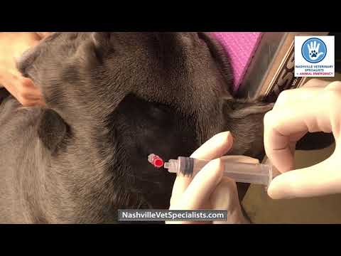
The most common cause of superficial vein inflammation in dogs encountered in clinical practice is irritation from intravenous catheter placement.
IV catheters are essential tools in veterinary medicine. They allow fluid therapy, medication administration, and anaesthetic delivery during procedures and hospital stays. But any foreign object placed within a vein creates a degree of irritation to the vessel wall. When a catheter remains in place for an extended period, when it is not maintained with strict hygiene protocols, or when the medications delivered through it are chemically irritating to vascular tissue, the inflammatory response can escalate beyond normal.
Hypertonic solutions, certain antibiotics, chemotherapy agents, and other medications with high osmolarity or low pH are particularly prone to causing chemical irritation to the vein wall when delivered through a catheter.
Poor aseptic technique during catheter placement or maintenance introduces bacteria directly into the vessel, creating conditions for septic thrombophlebitis, which is significantly more serious than sterile inflammation.
Infection or Injury
Beyond catheter-related causes, superficial vein inflammation can develop from direct bacterial infection of the vein or surrounding tissue following skin wounds, bites, or puncture injuries near a superficial vessel.
Bacteria that colonise a wound adjacent to a vein can spread to the vessel wall directly or through the lymphatic system. The most commonly implicated organisms in canine superficial vein infections are Staphylococcus species. Understanding conditions like staph infection in dogs provides important context for how bacterial infections can complicate localised tissue injury and why antibiotic selection matters in treatment.
Blunt trauma to a limb can damage a vein directly, triggering an inflammatory response without any infectious component. Post-traumatic phlebitis generally resolves more readily than infected phlebitis, as the primary driver is tissue damage rather than an active infection requiring antibiotic treatment.
Underlying Health Conditions
Certain systemic conditions create an environment in which superficial vein inflammation is more likely to develop or more difficult to resolve.
Obesity impairs circulation, particularly in the limbs, reducing the efficiency of venous return and creating localised areas of poor perfusion that are more susceptible to inflammatory change.
Immune system disorders reduce the body’s ability to contain and resolve inflammation and infection effectively. Dogs on immunosuppressive medications are at elevated risk of catheter-related infections.
Hypercoagulable states, conditions in which the blood clots more readily than normal, increase the risk of clot formation within veins following even minor irritation. Certain cancers, protein-losing diseases, and adrenal disorders can produce hypercoagulable states in dogs.
Chronic diseases that reduce overall vascular health, including diabetes mellitus and conditions causing prolonged inflammation, increase vulnerability to vein-related complications.
Diagnosing Superficial Vein Inflammation in Dogs
Veterinary Examination
Diagnosis of superficial thrombophlebitis typically begins with the physical examination findings described in the symptoms section. A veterinarian will assess the affected limb by palpating the vein and surrounding tissue to identify warmth, firmness, swelling, and pain.
The examination also includes an assessment of the dog’s overall condition, temperature, and any systemic signs that might suggest the infection has spread beyond the localised area.
The history of recent IV catheter placement, injections, or trauma to the area provides important context that helps the veterinarian narrow the likely cause quickly.
Blood Flow and Imaging Tests
Doppler ultrasound is the most useful imaging tool for evaluating suspected phlebitis. It visualises the blood vessel, detects abnormal flow patterns caused by clot formation, assesses the degree of vessel wall thickening, and can identify whether a clot is present within the lumen. Doppler imaging is non-invasive, quick to perform, and provides clinically actionable information about the extent of the condition.
Standard X-rays are of limited value for diagnosing phlebitis directly but may be used to evaluate the surrounding bone and tissue if deeper injury is suspected, particularly in cases involving trauma near a joint. The relationship between vascular and musculoskeletal injury in traumatic cases may also have implications for conditions like osteochondritis dissecans in dogs, where joint and connective tissue involvement requires comprehensive evaluation.
Blood cultures are taken when septic thrombophlebitis is suspected, to identify the causative organism and guide antibiotic selection. A blood culture from a dog with systemic signs of infection provides definitive information about which bacteria are present and which antibiotics will be most effective against them.
Complete blood count and biochemistry panel assess the degree of systemic inflammation and infection, evaluate organ function, and provide baseline data for monitoring treatment response.
Treatment for Superficial Vein Inflammation in Dogs
Removing Catheters or Irritants
The first and most important step when phlebitis is related to an IV catheter is to remove the catheter immediately. Continuing to use an inflamed vein as a catheter site allows the irritation and potential infection to worsen. The catheter is removed, the site is cleaned, and if continued IV access is required, a fresh catheter is placed in a different location on a different limb.
For mild, sterile phlebitis caused purely by catheter irritation without infection, this step alone, combined with supportive care, is often sufficient for resolution.
When chemical irritation from a specific medication is identified as the cause, reformulating the delivery method where possible, diluting the solution, or switching to an alternative route of administration addresses the underlying trigger.
Antibiotics and Anti-Inflammatories
When bacterial infection is confirmed or strongly suspected, antibiotic therapy is essential. The choice of antibiotic is ideally guided by culture and sensitivity testing, which identifies the specific organism and the drugs it responds to. While awaiting culture results, broad-spectrum antibiotics covering the most likely organisms are started.
The duration of antibiotic treatment depends on the severity of the infection. Mild superficial infections may be treated for seven to fourteen days. More extensive or deep infections require longer courses and close monitoring for resolution.
Non-steroidal anti-inflammatory medications reduce pain and local inflammation, improving the dog’s comfort during recovery. They are used as part of the overall pain management approach rather than as a standalone treatment.
Local Therapy and Wound Care
Warm compresses applied gently to the affected area several times daily improve local circulation, reduce discomfort, and can help encourage resolution of the localised inflammatory reaction. The compress should be warm but not hot, applied for ten to fifteen minutes at a time.
Wound care at the catheter site or any area of skin breakdown involves gentle cleaning with appropriate antiseptic solutions, removal of any discharge or crust, and dressing as needed to protect the area from further contamination.
Restricted activity during the acute phase prevents additional trauma to the affected limb and allows the inflammatory process to resolve without ongoing mechanical disruption.
Hydrotherapy and gentle physiotherapy may be introduced as the acute phase resolves to promote circulation and tissue healing, particularly in dogs where the inflammation has caused significant surrounding tissue involvement.
Related Products


Prognosis for Dogs with Phlebitis
For mild to moderate superficial phlebitis without significant infection, the prognosis is generally very good. Once the irritating source is removed and appropriate treatment is initiated, the inflammatory process typically resolves over days to a few weeks. The affected vein may remain slightly firm for a longer period as the tissue heals, but functional recovery is usually complete.
For septic phlebitis, the prognosis is good when treatment is started promptly, and the infection is confined to the superficial vein and surrounding tissue. Antibiotic treatment guided by culture results, combined with local wound management, resolves most cases fully.
For cases where infection spreads to deeper tissues, involves multiple sites, or occurs in dogs with compromised immune function, the prognosis becomes more guarded, and recovery may require more prolonged treatment and monitoring.
The rare complication of clot dislodgement and pulmonary embolism changes the clinical picture significantly and requires urgent intensive management.
Monitoring Vein Health in Dogs
After any period of hospitalisation during which IV catheter access was used, owners should check the catheter site and the vein along which the catheter was placed for the first week after returning home.
Look for any redness, swelling, warmth, or firmness developing at or near the site. Note whether your dog is favouring the affected leg, licking the area excessively, or showing signs of pain on handling.
If any of these signs appear, contact your veterinarian promptly rather than waiting to see if they resolve independently. Early intervention for developing phlebitis is faster, simpler, and less expensive than treating an established infection.
For dogs with known health conditions that increase their risk of vein inflammation or clot formation, discussing a monitoring plan with your veterinarian before hospitalisation is a practical and worthwhile step.
Preventing Superficial Vein Inflammation
Prevention of catheter-related phlebitis centres on strict aseptic technique and appropriate catheter management throughout the period of use.
Sterile insertion technique is the most important preventive measure. Every IV catheter should be placed using sterile equipment and proper skin preparation to minimise bacterial introduction at insertion.
Regular catheter site inspection during hospitalisation allows nursing staff to detect early signs of phlebitis before they progress. Any redness, swelling, or discharge at a catheter site should prompt immediate catheter removal and site assessment.
Scheduled catheter replacement within recommended timeframes reduces the cumulative irritation that leads to chemical and mechanical phlebitis. No catheter should remain in place longer than necessary or beyond the recommended maximum duration.
Minimising use of irritating solutions through dilution where clinically acceptable, using appropriate infusion rates, and choosing less irritating delivery routes when alternatives exist all reduce the chemical trauma to the vein wall.
At home, keeping wounds, bite injuries, and skin lesions clean and monitored reduces the risk of secondary bacterial infection reaching superficial veins.
Early Treatment Leads to Full Recovery
Superficial vein inflammation is not a dramatic condition in most cases. It does not typically threaten life. It does not require emergency intervention in its mild forms.
But it does require attention.
A warm, firm, swollen area along your dog’s leg following a vet visit is your signal to act. Do not wait for it to worsen. Do not assume it will resolve on its own without assessment. Contact your veterinarian, describe what you are seeing, and follow their guidance.
The dogs who recover quickly from phlebitis are the ones whose owners noticed the early signs and acted on them within a day or two. Caught early, this condition is simple to treat and resolves completely. Left to progress, it becomes more complicated, more painful, and more costly to manage.
VOSD is committed to giving dog owners the knowledge they need to recognise health changes early and respond appropriately. For more on the full range of conditions that affect dogs and how to identify them, the broader VOSD veterinary resource library provides detailed, clinically grounded guidance on canine health across every body system.
Your dog’s health between vet visits is in your hands. And informed hands make the best caregivers.


