

When the nerve that carries vision to the brain becomes inflamed, sight can disappear overnight.
Not gradually, the way it does in progressive retinal atrophy. Not with the warning signs of redness and squinting that accompany a corneal ulcer or uveitis. With optic neuritis, a dog can go from apparently normal vision to complete blindness within hours, sometimes within a single day, and the eye itself may look entirely normal from the outside.
This is what makes optic neuritis one of the most alarming eye conditions in veterinary medicine. The problem is not in the eye. It is in the nerve that connects the eye to the brain. And because that nerve is part of the central nervous system, optic neuritis is not simply an eye problem. In many cases, it is a neurological emergency that reflects serious disease elsewhere in the body or the brain.
Sudden vision loss in a dog is always an emergency. Optic neuritis is one of the most important reasons why.
What is Optic Nerve Swelling (Optic Neuritis) in Dogs?
The optic nerve is the second cranial nerve. It carries the electrical signals generated by the photoreceptor cells of the retina directly to the visual centres of the brain. Without an intact, functioning optic nerve, the brain receives no visual information, regardless of how healthy the eye itself is.
Optic neuritis is inflammation of the optic nerve. When the nerve becomes inflamed, the swelling disrupts the transmission of visual signals along its length. Depending on the severity of the inflammation, vision may be partially impaired or completely lost. The inflammation damages the myelin sheath that insulates the nerve fibres and, in severe or prolonged cases, the nerve fibres themselves.
Because the optic nerve is considered part of the central nervous system rather than the peripheral nervous system, its anatomy and biology are fundamentally different from those of other nerves in the body. Inflammation here is not simply local. It frequently reflects a broader disease affecting the brain and spinal cord, which is why optic neuritis demands a systemic investigation rather than an isolated eye treatment.
The condition is most commonly seen as a secondary manifestation of underlying systemic or neurological disease. True primary optic neuritis, where the nerve is inflamed without any identifiable underlying cause, is considered less common and is diagnosed only after all other causes have been excluded.
Symptoms of Optic Nerve Swelling in Dogs
The clinical picture of optic neuritis is dominated by visual loss, which is often sudden and severe.
Sudden Blindness
Acute onset of blindness is the hallmark presenting sign. A dog that was navigating its environment normally may suddenly walk into walls, furniture, and doorways. It may fail to respond to objects approaching its face, stop tracking moving toys or people, or appear completely disoriented in spaces it knows well.
The speed of onset is diagnostically important. Gradual vision loss over weeks suggests a degenerative or slowly progressive condition. Vision loss that develops over hours to a couple of days is a red flag for optic neuritis or other acute neurological events.
Pupillary Light Response Abnormalities
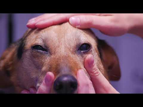
In a normal dog, shining a light into the eye causes both pupils to constrict. In optic neuritis, the affected eye or eyes show a reduced or absent pupillary light response because the nerve carrying the signal from the retina to the brain is not functioning properly. The pupils may appear persistently dilated, fixed, or slow to respond even in bright light.
This sign is one of the most clinically useful indicators of optic nerve dysfunction and is a key part of the neurological eye examination.
Behavioural and Navigational Signs
- Bumping into objects that were previously avoided without difficulty
- Hesitation at doorways, steps, and transitions between surfaces
- Apparent confusion or disorientation, particularly in unfamiliar environments
- Reluctance to move, particularly in low light
- Startling when approached, as the dog did not see the person or object coming
Neurological Signs
Because optic neuritis is frequently associated with broader central nervous system disease, some dogs present with signs that extend beyond vision loss. Seizures, changes in behaviour or personality, head tilt, circling, altered gait, or other neurological abnormalities alongside sudden blindness strongly suggest that the problem extends to the brain itself and makes the diagnostic urgency even greater.
Causes of Optic Nerve Swelling in Dogs
Immune-Mediated Disease
Immune-mediated optic neuritis, where the dog’s own immune system attacks the optic nerve tissue, is among the most commonly identified causes. This may occur as an isolated condition or as part of a broader immune-mediated meningoencephalitis, inflammation of the brain and its surrounding membranes. Granulomatous meningoencephalomyelitis, commonly abbreviated as GME, is a specific immune-mediated inflammatory brain disease in dogs that frequently involves the optic nerves and is an important cause of acute blindness in middle-aged small breed dogs.
Infectious Causes
Several infectious agents can cause optic neuritis by directly infecting the nerve tissue or by producing inflammatory responses that damage it.
- Fungal infections including cryptococcosis and aspergillosis, which are particularly relevant in immunocompromised dogs and can spread from the respiratory system to the central nervous system. Our guide on Parasitic Infection of the Respiratory Tract in Dogs provides relevant background on how respiratory infections can have systemic and neurological implications.
- Canine distemper virus, which has a specific tropism for nervous tissue and is a recognised cause of optic neuritis, particularly in unvaccinated dogs
- Tick-borne diseases including ehrlichiosis and Rocky Mountain spotted fever
- Protozoal infections, including toxoplasmosis and neosporosis, which can invade nervous tissue and produce optic nerve inflammation
Neoplasia
Tumours affecting the optic nerve directly, such as optic nerve meningiomas, or tumours involving the brain, orbit, or nasal cavity that compress or invade the optic nerve, can produce signs of optic neuritis. Systemic cancers, including lymphoma, can also infiltrate the optic nerve as part of their spread.
Toxins
Toxic optic neuropathy, where exposure to certain substances causes optic nerve damage, is a less common but recognised cause. Lead toxicity is the best documented toxic cause of optic nerve swelling in dogs. Metabolic toxins accumulating in severe systemic disease, including renal failure, can also affect nervous tissue. Our guide on Kidney Filtration Problems in Dogs discusses how advanced renal disease can produce systemic complications affecting multiple organ systems including the nervous system.
Related Videos


The Neurological Link: Why the Optic Nerve Is Not Just an Eye Problem
The optic nerve is a direct extension of the brain. It is surrounded by the same protective membranes, the meninges, that surround the brain and spinal cord, and it shares the same cerebrospinal fluid environment.
This anatomy means that any disease process affecting the meninges, the brain, or the cerebrospinal fluid can directly affect the optic nerve. Meningitis, encephalitis, and meningoencephalomyelitis all produce conditions in which the optic nerve is inflamed alongside the broader central nervous system.
The clinical implication of this is critical: a dog presenting with optic neuritis must be evaluated not just for eye disease but for brain disease. The vision loss may be the most visible sign of a condition that is simultaneously affecting other parts of the central nervous system. Neurological examination, advanced imaging, and cerebrospinal fluid analysis are often necessary parts of the diagnostic process.
How Optic Neuritis Develops
Inflammation of the optic nerve begins when the triggering cause, whether immune-mediated, infectious, neoplastic, or toxic, produces an inflammatory response within or around the nerve tissue.
The inflammatory cells and mediators that accumulate within the confined space of the optic nerve sheath cause localised swelling. This swelling compresses the nerve fibres and disrupts the myelin sheath that normally allows rapid, efficient signal transmission. As the myelin is damaged, the speed and fidelity of signal transmission from the retina to the brain deteriorate. Partial damage produces reduced or impaired vision. Complete disruption of signal transmission produces blindness.
If the inflammation is controlled quickly enough, the myelin can partially regenerate, and visual function can recover. If the inflammation persists or the nerve fibres themselves are damaged beyond the myelin, the loss of function becomes permanent. This is why time is the single most critical variable in the prognosis of optic neuritis. The window for preserving vision is real, and it is not unlimited.
Types of Optic Nerve Swelling
Primary optic neuritis refers to cases where inflammation of the optic nerve occurs without an identifiable underlying cause. This is relatively uncommon in dogs and is a diagnosis of exclusion, made only after thorough investigation has failed to reveal an underlying disease.
Secondary optic neuritis is far more common and occurs as a consequence of systemic disease, central nervous system inflammation, infection, neoplasia, or toxic exposure. Treatment of secondary optic neuritis centres on identifying and addressing the underlying cause alongside managing the nerve inflammation.
Understanding which type is present directly determines the treatment approach and the prognosis for visual recovery. For a detailed look at a related condition involving pressure-related changes to the optic disc, our guide on Swelling of the Optic Disk in the Retina of Dogs provides important context on how papilledema differs from true inflammatory optic neuritis.
Diagnosis of Optic Nerve Swelling in Dogs
Ophthalmoscopic Examination
Fundoscopic examination of the retina allows direct visualisation of the optic disc, the point at which the optic nerve enters the eye. In optic neuritis, the disc appears swollen, raised, and often hyperaemic with indistinct margins. The retinal blood vessels adjacent to the disc may appear congested or abnormal. This finding, combined with the clinical signs of vision loss and poor pupillary response, provides the initial clinical diagnosis.
However, it is important to note that in some cases of optic neuritis, particularly those affecting the nerve behind the eye rather than at the disc, the fundus appearance may be normal despite significant nerve dysfunction. A normal-looking optic disc does not exclude optic neuritis.
Neurological Examination
A full neurological examination assesses cranial nerve function, proprioception, reflexes, gait, and mental status. Abnormalities on neurological examination alongside optic neuritis strongly suggest central nervous system involvement and escalate the urgency of the investigation.
Advanced Imaging
MRI of the brain and orbits is the gold standard imaging investigation for optic neuritis. It allows visualisation of the optic nerves, the optic chiasm, the brain parenchyma, and the meninges, identifying areas of inflammation, oedema, or masses that explain the nerve swelling. CT imaging provides less soft tissue detail but may be available when MRI is not.
Cerebrospinal Fluid Analysis
In cases where central nervous system inflammation is suspected, analysis of cerebrospinal fluid obtained by lumbar puncture provides direct information about the inflammatory cell profile and whether infection or immune-mediated disease is driving the process.
Systemic Blood and Urine Testing
Infectious disease panels, complete blood count, biochemistry, and urinalysis are standard components of the workup, identifying systemic infections, metabolic disease, or organ dysfunction that may be contributing to the neurological presentation.
Treatment of Optic Nerve Swelling in Dogs
Treating the Underlying Cause
This is the most important principle. Optic neuritis caused by a specific infection requires appropriate antimicrobial treatment, whether antifungal agents, antibiotics, or antiprotozoal medication, based on the identified organism. Optic neuritis caused by a compressive mass may require surgical or oncological management. Immune-mediated optic neuritis requires immunosuppressive therapy.
Without addressing the underlying cause, anti-inflammatory treatment alone provides temporary relief while the driving disease continues to progress.
Anti-Inflammatory and Immunosuppressive Therapy
Corticosteroids are the primary anti-inflammatory agents used in the management of optic neuritis, particularly in immune-mediated cases. High doses are typically used initially to rapidly reduce the inflammation compressing the nerve fibres, with gradual tapering based on the dog’s response. In some cases, additional immunosuppressive agents are added for long-term management of immune-mediated disease.
Antimicrobial Treatment
Where infectious causes are identified or strongly suspected, appropriate antimicrobial therapy is initiated simultaneously with anti-inflammatory treatment. The specific agents depend on the identified or suspected organism.
Supportive Care
Dogs that have lost vision require supportive management during the treatment period and beyond. Keeping the environment consistent, avoiding rearranging furniture, using verbal cues to guide the dog, and preventing access to hazards are practical measures that reduce the risk of injury during the period of visual impairment.
Related Products

Prognosis and Recovery
The prognosis for optic neuritis varies considerably depending on the underlying cause, the severity of nerve damage at the time treatment is initiated, and how quickly treatment is started.
Dogs with immune-mediated optic neuritis that is identified and treated promptly have a reasonable chance of partial or full visual recovery as the inflammation resolves and the myelin regenerates. Recovery, when it occurs, typically begins within days to weeks of initiating treatment and may continue for several weeks.
Dogs with optic neuritis caused by infectious diseases have a prognosis that depends on how effectively the infection can be controlled. Some infectious causes, including certain fungal infections, carry a guarded prognosis for both vision and overall survival.
When nerve fibre damage has progressed to optic nerve atrophy, the structural damage is permanent, and vision cannot be recovered. This is why every day without treatment represents a reduction in the probability of visual recovery.
Some dogs treated successfully for optic neuritis experience recurrence when medication is reduced or discontinued, particularly in immune-mediated cases. Long-term monitoring and maintenance therapy may be required.
Complications if Left Untreated
Optic neuritis that is not treated progresses to outcomes that cannot be reversed.
- Permanent blindness from optic nerve fibre death beyond the point of recovery
- Optic nerve atrophy, where the nerve physically degenerates, and the optic disc appears pale and shrunken on fundoscopy
- Progressive neurological disease, if an underlying central nervous system condition is not addressed, potentially affecting other neurological functions beyond vision
- Seizures and other neurological deficits if the broader central nervous system inflammatory process advances unchecked
The eye may look normal throughout. From the outside, there is nothing to indicate that a catastrophic process is destroying one of the most critical structures in the visual system. This is precisely why sudden vision loss demands immediate veterinary evaluation rather than observation.
Is Optic Nerve Swelling an Emergency?
Yes. Without qualification.
Sudden vision loss in a dog, of any degree, is a neurological and ophthalmological emergency. The window for preserving visual function in optic neuritis is narrow. The underlying causes are serious. The complications of delayed treatment are irreversible.
A dog that has suddenly lost vision, shows poor or absent pupillary light responses, or is displaying confusion and disorientation alongside visual changes needs to be seen by a veterinarian on the same day. Emergency referral to a facility with neurology or ophthalmology capability is likely to be necessary.
Do not wait to see if it improves on its own. It will not.
Optic Neuritis vs Papilledema: An Important Distinction
Both conditions produce a swollen-appearing optic disc on fundoscopic examination, which is why they can be difficult to distinguish clinically without additional investigation.
Optic neuritis is inflammation of the optic nerve itself. The swelling is caused by inflammatory cells and oedema within the nerve tissue. Visual function is typically significantly impaired because the inflammation disrupts signal transmission.
Papilledema is swelling of the optic disc caused by elevated intracranial pressure, where increased pressure within the skull is transmitted along the cerebrospinal fluid pathway surrounding the optic nerve to the optic disc. In papilledema, visual function may be relatively preserved initially because the nerve fibres themselves are not directly inflamed, only mechanically displaced by the pressure. The treatment focus in papilledema is reducing intracranial pressure rather than suppressing nerve inflammation.
Distinguishing between the two requires consideration of the full clinical picture, visual function testing, neurological examination, and advanced imaging. The distinction matters because the underlying causes and treatment approaches differ significantly.
When to See a Vet
Seek emergency veterinary assessment immediately if your dog shows:
- Sudden loss of vision or any acute deterioration in the ability to navigate
- Pupils that appear fixed, dilated, or unresponsive to light
- Bumping into objects that were previously avoided
- Disorientation or confusion, particularly if onset is acute
- Any combination of vision change and neurological signs, including seizures, altered behaviour, or changes in gait
Do not wait. Do not monitor overnight. Vision loss from optic neuritis can become permanent within days of onset, and the underlying causes are serious enough to require immediate professional evaluation regardless of what other signs are or are not present.


