

Any lump or growth inside a dog’s mouth deserves immediate attention. Not next month. Not at the next routine appointment. Now.
Oral masses in dogs range from entirely benign growths that require straightforward removal to aggressive cancers that invade bone, spread to lymph nodes, and require urgent, multimodal treatment. The two can look remarkably similar on the surface. A growth that appears slow-moving and painless can turn out to be a malignant tumour on biopsy. A mass that looks alarming may prove to be benign.
The only way to know is through proper diagnosis. And the earlier that diagnosis happens, the wider the treatment options available and the better the outcome for your dog.
What Are Oral Masses in Dogs?
Oral masses are abnormal growths that develop in the tissues of the mouth. They can arise from the gums, tongue, lips, palate, cheeks, or the bone of the jaw itself. They vary in size, texture, appearance, and behaviour depending on their cellular origin.
Some are benign, meaning they grow locally without invading surrounding structures or spreading to other parts of the body. Others are malignant, meaning they actively invade adjacent tissue, destroy bone, and in many cases metastasise to regional lymph nodes and the lungs.
The critical point is that no oral mass can be reliably characterised by appearance alone. Size, colour, surface texture, and rate of growth do not distinguish benign from malignant with sufficient accuracy to guide treatment decisions. A tissue biopsy is always required.
Types of Oral Masses in Dogs
Benign Oral Masses
Benign oral masses grow slowly, do not invade surrounding structures, and do not spread to other organs. They are nonetheless clinically significant because they can cause discomfort, disrupt normal eating and tooth alignment, and in some locations, interfere with breathing or swallowing.
Epulis, now more precisely called peripheral odontogenic fibroma, is one of the most common benign oral growths in dogs. It arises from the periodontal ligament tissue around the teeth and typically appears as a firm, smooth growth on the gum line. Ameloblastoma in dogs is another type of odontogenic tumour that, while often locally aggressive in its behaviour, is classified as benign in that it does not typically spread distantly.
Oral papillomas, caused by the canine papillomavirus, appear as multiple small, wart-like growths, most commonly in young dogs, and frequently resolve on their own as the immune system responds.
Malignant Oral Tumours
Malignant oral tumours are the most serious category and require urgent diagnosis and treatment. The three most commonly diagnosed malignant oral tumours in dogs are malignant melanoma, squamous cell carcinoma, and fibrosarcoma.
Malignant melanoma is the most frequently diagnosed oral cancer in dogs and is one of the most aggressive. It spreads rapidly to the lymph nodes and lungs and can cause significant local bone destruction. Squamous cell carcinoma is locally invasive and deeply destructive to underlying bone, though it tends to spread to distant organs more slowly in the early stages. Fibrosarcoma is a tumour of the connective tissue that grows aggressively into surrounding structures and has a high local recurrence rate after surgery.
For a detailed overview of these conditions and their management, our guide to dog mouth cancer covers the full clinical picture.
Symptoms of Oral Masses in Dogs
Because the mouth is not an area most pet parents examine regularly, oral masses are frequently discovered late. Knowing the signs, both visible and behavioural, significantly improves the chance of early detection.
Common Clinical Signs
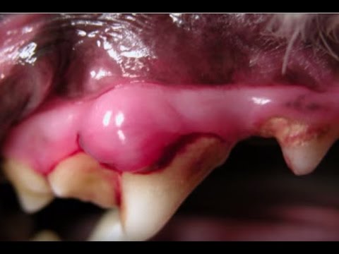
- A visible lump, swelling, or irregular tissue growth on the gums, tongue, lips, or palate
- Severely bad breath, often markedly worse than typical dental halitosis
- Drooling, which may be blood-tinged
- Bleeding from the mouth without an obvious injury
- Difficulty picking up food, chewing, or swallowing
- Loose teeth without a clear dental disease explanation
- Facial swelling, particularly around the jaw, cheek, or below the eye
- Unexplained weight loss as eating becomes progressively more difficult
Extremely foul breath is frequently the first sign that draws a pet parent’s attention. While bad breath in dogs is common and usually attributable to dental disease, a sudden worsening or an unusually severe odour that does not improve with dental treatment warrants a thorough oral examination including assessment for mass lesions.
Related Videos


Causes of Oral Masses in Dogs
The causes of oral tumours in dogs are not fully understood, and in many cases no single identifiable trigger exists. Several risk factors have been documented.
Genetic and Breed Factors
Certain breeds have a documented higher incidence of oral tumours. Golden Retrievers, Boxers, German Shepherds, Cocker Spaniels, and Poodles appear more frequently in oral tumour statistics than the general dog population. Understanding general dog tumour risk across breeds provides useful context for pet parents of predisposed dogs.
Breed predisposition does not make oral cancer inevitable. It does make routine oral examinations a particularly important part of preventive care.
Age and Cellular Changes
Oral tumours are significantly more common in middle-aged to older dogs. The cumulative cellular changes that occur over time, including DNA damage, reduced immune surveillance, and the effects of chronic low-grade inflammation, all increase the likelihood of abnormal cell proliferation as dogs age.
This is not a reason to be complacent in younger dogs, as oral masses do occur across all age groups, but it does make regular oral examination especially important in dogs over seven years of age.
Chronic Inflammation or Injury
Persistent oral inflammation, chronic periodontal disease, and repeated physical irritation of the oral mucosa are all associated with an increased risk of abnormal tissue changes over time. The mechanism involves repeated cycles of cellular damage and repair, which over time can increase the probability of mutations that lead to tumour development.
Environmental and Unknown Factors
In many cases, no clear cause can be identified. Some studies suggest associations with environmental toxin exposure and second-hand smoke, but oral tumours frequently develop in dogs with no identifiable risk factor. This reinforces the value of routine examination rather than waiting for risk factors to be present before examining the mouth.
How Veterinarians Diagnose Oral Masses in Dogs
Oral Examination
The starting point is a thorough visual and manual examination of the entire mouth under general anaesthesia. The vet assesses the size, location, surface characteristics, and texture of the mass, and palpates the regional lymph nodes for enlargement. The extent of any involvement of the jaw bone is noted.
General anaesthesia is necessary for a complete and accurate examination. Attempting to assess oral masses in a fully conscious dog frequently results in an incomplete picture of the mass and its margins.
Biopsy (Critical Step)
Biopsy is the single most important diagnostic step and should never be skipped or delayed. A tissue sample from the mass is sent for histopathological analysis, which identifies the cell type, confirms whether the growth is benign or malignant, and in the case of malignant tumours, characterises the grade and likely behaviour of the cancer.
No treatment should be planned without biopsy confirmation. A mass that looks benign may not be, and the treatment for a benign epulis is entirely different from the treatment for a malignant melanoma.
Imaging (X-ray, CT, and MRI)
Following confirmation of a malignant tumour, imaging is essential for staging. Dental radiographs assess local bone involvement. CT scanning of the head and neck provides the most detailed information about tumour extent, bone destruction, and regional lymph node involvement, and is used for surgical planning. Chest X-rays or CT assess for pulmonary metastasis, which significantly influences the treatment approach and prognosis.
Treatment for Oral Masses in Dogs
Surgical Removal (Primary Treatment)
Surgery is the primary treatment for most oral masses, both benign and malignant. For benign masses, complete excision with clear margins typically results in cure or long-term remission. For malignant tumours, surgery aims to remove the mass with the widest possible margins, which in many cases requires removal of a portion of the jaw bone.
Dogs adapt remarkably well to partial mandibulectomy or maxillectomy. Most resume eating comfortably within weeks of recovery, and the improvement in quality of life after removal of a painful or infected mass is typically significant and rapid.
Radiation Therapy and Chemotherapy
Radiation therapy is used when surgery cannot achieve complete removal, when the tumour is located in a position that limits surgical margins, or as an adjunct to surgery to reduce the risk of local recurrence. Its effectiveness varies by tumour type, with squamous cell carcinoma responding more reliably than malignant melanoma.
Chemotherapy plays a more limited role in oral tumours than in some other canine cancers, but may be used as part of a multimodal approach for malignant melanoma and other systemic cancers. Immunotherapy, including a licensed melanoma vaccine for dogs, is also available at specialist centres for eligible cases.
Supportive and Palliative Care
When curative treatment is not pursued or not possible, the focus shifts to managing pain, maintaining nutrition, and preserving quality of life. This is a valid and compassionate choice, and skilled palliative management can provide meaningful comfort for extended periods. Anti-inflammatory medications, pain relief, appetite stimulants, and soft dietary modifications are all part of palliative care planning.
Related Products


Prognosis and Life Expectancy
Prognosis varies considerably depending on the tumour type, location, size, and stage at diagnosis.
Benign masses carry an excellent prognosis following complete surgical removal. Recurrence is uncommon when adequate margins are achieved.
Malignant tumours carry a more variable prognosis. Rostral (front of mouth) tumours that can be removed with clean margins have significantly better outcomes than caudal (back of mouth) tumours where surgical access is limited. Early-stage tumours without lymph node or lung involvement respond better to treatment than those diagnosed after metastasis has occurred.
The consistent finding across all oral tumour types is that earlier diagnosis and treatment produces better outcomes. A mass identified at a small, localised stage offers fundamentally more treatment options than one discovered after significant bone invasion or distant spread.
Complications of Untreated Oral Masses
Infection and Ulceration
As oral masses grow, their surface frequently breaks down, ulcerates, and becomes infected. This causes persistent pain, bleeding, and severe halitosis that significantly affects the dog’s quality of life and makes eating increasingly distressing.
Difficulty Eating and Weight Loss
Oral pain, mechanical obstruction from the growing mass, and the disruption to normal swallowing mechanics all progressively reduce a dog’s ability to eat comfortably. Weight loss follows, further weakening the dog’s overall condition and resilience.
Spread to Other Organs (Metastasis)
Malignant oral tumours, particularly malignant melanoma, spread to the regional lymph nodes and lungs. Once metastasis is established, treatment becomes significantly more complex and the prognosis more guarded. This is the most compelling reason why early biopsy and staging matter so profoundly in oral tumour management.
When to See a Veterinarian
Contact your veterinarian immediately if you notice any of the following in your dog:
- Any visible lump, bump, or abnormal tissue in the mouth
- Bleeding from the mouth without an obvious injury
- Suddenly severe bad breath that does not respond to dental care
- Difficulty eating, dropping food, or reluctance to chew
- Facial swelling, particularly around the jaw
- Loose teeth without a clear dental explanation
Make oral examination a regular part of your handling and grooming routine. Lift the lips, look at the gums, cheeks, and tongue. A mass found small is a problem with options. A mass found large is a problem with fewer.


