


A maggot wound is not simply an infected wound. It is active, ongoing tissue destruction happening in real time.
When flies lay eggs in a warm, moist, or already-injured area of a dog’s skin, those eggs hatch into larvae within hours. The larvae burrow into tissue, feeding on it as they go. They create deep tunnels beneath the skin surface. They release secretions that break down healthy tissue to make it accessible. The wound that was visible on the surface is often the smallest part of what is happening underneath.
This is myiasis, the infestation of living tissue by fly larvae, and it is one of the most urgent wound conditions a dog can develop. It is also one of the most preventable. And it is almost always a sign that a dog has been suffering without adequate care or observation.
What Happens in a Maggot Wound
The progression from fly eggs to serious tissue destruction is rapid and follows a predictable sequence.
A fly, typically a blowfly or screwfly, is attracted to moisture, discharge, odour, or the warmth of an open wound. It lands and lays clusters of eggs at or near the wound margin. Under warm conditions, those eggs can hatch into first-stage larvae within eight to twelve hours.
The larvae immediately begin burrowing. They move away from light and toward warmth, which means they go deeper into the tissue rather than remaining at the surface. As they feed, they release proteolytic enzymes that liquefy the surrounding tissue, creating a progressively enlarging cavity of necrotic material that provides further nutrition and habitat.
Within twenty-four to forty-eight hours of egg laying, what may have appeared to be a manageable surface wound can become a large, deep cavity with hundreds of larvae, extensive tissue death, and rapidly spreading bacterial infection. The foul odour associated with myiasis comes from the combination of tissue necrosis and bacterial overgrowth in the warm, moist environment that the larvae create.
Left untreated, the infection extends into deeper tissue layers, enters the bloodstream, and produces septic shock. The dog deteriorates from local wound disease to systemic crisis quickly.
Symptoms of Maggot Wounds in Dogs
Symptoms follow the progression of the infestation from surface to deep involvement.
Early signs include a wound or area of skin that appears moist or irritated, often with the dog attending to it by licking or biting. There may be a faint odour. The area may appear slightly swollen. At this stage, the larvae may not yet be visible, particularly if the infestation began in a hair-covered area.
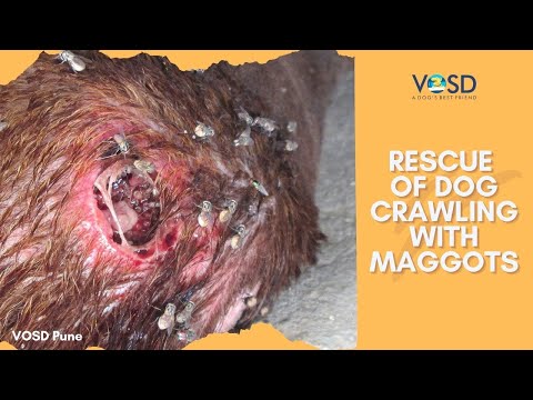
Moderate signs include a distinctly foul, sweet-rotten odour from the wound, visible small white or cream-coloured larvae moving within the wound, a wet or weeping wound surface that appears to be enlarging despite no additional trauma, and increasingly distressed behaviour from the dog, including constant licking, restlessness, and sensitivity around the area.
Severe signs indicate deep tissue involvement and systemic effects. The wound is large, deep, and may reveal subcutaneous tunnels when the hair is parted. The tissue at the wound margins appears grey, black, or otherwise necrotic. The dog is lethargic, may be febrile, is not eating, and shows signs of systemic illness. The sheer number of larvae present may be visible as movement within the wound, even from a distance.
The detailed guide to treating maggot wounds provides further specific guidance on wound management approaches and the range of presentations seen in clinical practice.
Why Maggot Wounds Happen
Myiasis is almost always preventable. Understanding the conditions that allow it to develop identifies where prevention needs to be directed.
Untreated wounds are the most straightforward cause. Any break in the skin, whether from injury, surgery, a bite wound, or pressure sore, that is not cleaned and monitored provides a landing site for flies. A wound that is left unobserved for even twenty-four hours in warm weather can develop an infestation.
Moisture from urine or faeces contamination is a major predisposing factor. Dogs that are incontinent, paralysed, or unable to move away from their waste develop perpetually damp skin around the hindquarters. Flies are powerfully attracted to this environment. Urine scalding and faecal contamination of the skin also damage the skin barrier, providing additional entry points for larvae.
Immobile or weakened dogs are at extreme risk. A dog that cannot groom itself, cannot move away from flies, or cannot respond to the irritation of early infestation is unable to protect itself in any way. Elderly dogs, paralysed dogs, and very sick dogs are among the most vulnerable.
Poor hygiene and inadequate wound observation in both owned and stray dogs allows infestations to develop and progress to severity before they are noticed. In warm, humid climates, a wound can go from clean to severely infested within a single day of inattention.
Post-surgical neglect is a specific clinical context. Surgical wounds that are not kept clean, that get wet, or that the dog is allowed to lick excessively provide ideal fly-laying sites during the healing period.
Related Videos


Immediate First Aid for a Maggot Wound
This is the section where correct action has the most direct impact. Every step matters, and the sequence should be followed precisely.
Isolate the dog from other animals and from the fly-accessible environment. Move the dog indoors or to a clean, sheltered space where additional fly exposure cannot occur during your assessment and first aid.
Clip the hair around the wound thoroughly. Hair traps moisture, hides larvae, and prevents effective cleaning. Using scissors or clippers, remove as much hair as possible from the wound margins and the surrounding area. This is essential before any cleaning or removal attempt.
Flush the wound with an antiseptic solution. A dilute chlorhexidine or povidone-iodine solution applied generously to the wound helps kill surface larvae, removes loose material, and begins to clean the wound bed. Use a syringe to direct the flow into the wound with some pressure, which helps dislodge larvae from the surface.
Remove visible surface larvae carefully. Using blunt-ended tweezers or forceps, remove individual larvae that are visible at the surface and wound margins. Work methodically. Place removed larvae into a sealed container or bag to prevent reinfestation.
Do not crush or break larvae during removal. This is critically important. Broken larvae release their gut contents and secretions directly into the wound tissue. These substances are toxic to the surrounding tissue and significantly worsen local damage and infection. Remove larvae whole.
Do not attempt to excavate deep tissue to retrieve larvae that are not visible. Pushing instruments into wound tunnels risks additional trauma and pushing larvae deeper. Surface removal is the limit of appropriate first aid. Deep removal is a veterinary procedure.
Apply a clean, non-stick antiseptic dressing loosely over the wound to protect it during transport.
Transport to a veterinary facility immediately. First aid reduces the immediate burden but does not address the full infestation, the deep tissue damage, or the systemic effects. No maggot wound can be fully managed at home.
What Not to Do
Several approaches that seem logical cause additional harm.
Do not pour random household chemicals into the wound. Turpentine, kerosene, petrol, undiluted bleach, and other harsh substances damage viable tissue as severely as they damage larvae. They worsen the wound, extend the area of necrosis, and make subsequent veterinary treatment more difficult.
Do not leave any visible larvae untreated while waiting for the vet. Even an hour of delay allows remaining larvae to burrow deeper and cause additional damage. Surface removal during the waiting period reduces the ongoing burden.
Do not ignore areas adjacent to the main wound. Larvae migrate. A dog with myiasis in one visible location may have larvae in surrounding tissue that are not yet visible. The entire surrounding area needs examination.
Do not delay veterinary care based on the wound appearing improved after first aid. Surface cleaning and partial larval removal do not address the deep components of the infestation or the secondary infection that has already begun.
How Veterinarians Diagnose a Maggot Wound
Diagnosis of myiasis is typically visual. The presence of larvae in a wound is confirmed on physical examination. The more important clinical work is assessing the extent of the infestation and the degree of systemic effect.
The veterinarian will assess the depth and size of the wound, identify all areas of larval activity, including tunnels extending beyond the primary wound, evaluate the viability of the surrounding tissue, and determine whether the dog is systemically affected through temperature, heart rate, blood pressure, and blood test findings.
Blood tests assess infection markers, kidney and liver function, and overall health status. Severely affected dogs with systemic illness require stabilisation before full wound management proceeds.
Treatment of Maggot Wounds in Dogs
| Step | Treatment |
|---|---|
| Remove larvae | Manual extraction under sedation |
| Clean wound | Antiseptic irrigation and debridement |
| Kill remaining larvae | Parasiticide application |
| Treat infection | Systemic antibiotics |
| Severe tissue loss | Surgical debridement or reconstruction |
Manual extraction of all larvae under sedation or anaesthesia is the primary treatment step. With the dog still and the wound fully accessible, the veterinary team methodically removes every larva from the wound and all extending tunnels. This is painstaking work because a single remaining larva can restart the infestation.
Debridement removes all necrotic and non-viable tissue from the wound. Tissue that has been destroyed by larval activity cannot heal and provides an ongoing infection substrate. Thorough debridement is essential, but must be balanced against preserving viable tissue needed for closure.
Antiseptic irrigation of the entire wound cavity follows debridement, flushing the wound with dilute antiseptic solution to reduce bacterial load.
Parasiticide application using dilute ivermectin or other approved antiparasitic agents applied directly to the wound kills any larvae that were not successfully extracted manually. This is used under veterinary guidance, not as a home treatment.
Systemic antibiotics are indicated in virtually all cases of significant myiasis because secondary bacterial infection is invariably present. The choice of antibiotic is guided by the degree of infection and, in severe cases, culture and sensitivity testing.
Wound management continues over subsequent days with dressing changes, reassessment for any missed larvae, and monitoring of healing. Some wounds require surgical closure once the infection is controlled. Others are managed as open wounds healing by secondary intention.
Pain management is an essential component of treatment. Myiasis wounds are painful, and appropriate analgesia supports the dog’s recovery and reduces the stress response that slows healing.
Related Products


Complications of Untreated Maggot Wounds
When myiasis is not treated promptly and aggressively, the consequences escalate beyond the local wound.
Deep tissue loss occurs as larvae consume progressively more tissue. Muscle, fascia, and in some locations, bone can be exposed. Large areas of tissue loss require extensive surgical reconstruction or result in permanent disfigurement.
Septic infection develops when the bacterial burden in the wound enters the bloodstream. The necrotic tissue created by myiasis is an ideal environment for anaerobic bacteria that produce particularly dangerous infections.
Toxaemia results from the systemic absorption of tissue breakdown products and bacterial toxins from the wound. Affected dogs develop fever, lethargy, reduced appetite, and deteriorating organ function.
Shock can develop from the combination of fluid loss through the wound, systemic infection, and pain. A dog in shock requires intensive resuscitation before wound management can proceed.
Death is the outcome of severe, untreated myiasis in a dog that does not receive veterinary intervention. It is not a rare outcome in cases where the infestation has been missed for several days in a dog that is already debilitated.
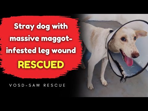
The difference between a wound that heals and one that becomes life-threatening is almost always the speed of intervention. Cases like the stray dog with a heart condition who found medical care and a home at VOSD show what is possible when dogs with severe, complex medical needs receive timely and expert care.
Preventing Maggot Wounds
Myiasis is almost entirely preventable with basic hygiene and observation.
Check your dog’s skin and coat daily, particularly during warm and humid months. Pay specific attention to less visible areas: under the tail, around the groin, between skin folds, and anywhere the dog has been licking persistently. Early detection before larvae are visible is the goal.
Clean any wound immediately. Any cut, scrape, bite wound, or skin break should be cleaned, assessed, and covered. Do not leave any open wounds exposed in warm weather without protection.
Keep the skin around the hindquarters clean and dry in dogs that are incontinent, elderly, or immobile. Urine and faeces contamination of skin must be cleaned promptly at every episode, not left until the next bathing session.
Control flies in the dog’s environment. Fly screens, fly traps, and avoiding leaving the dog in fly-accessible outdoor areas during peak fly activity periods reduce exposure. After any outdoor time in warm weather, check the dog’s coat for eggs, which appear as small clusters of white specks in the fur.
Groom regularly and thoroughly. Dense, matted, or dirty coats create microenvironments where moisture accumulates, and wounds can go unnoticed. Regular grooming maintains visibility and skin health.
Monitor post-surgical wounds carefully. Keep surgical sites clean, prevent licking with an Elizabethan collar if necessary, and check every day until fully healed.
Early Action Is the Difference
A maggot wound seen and treated on day one is a manageable wound. The same wound seen on day three is a surgical emergency. The difference is observation.
Every dog deserves the kind of attentive care that catches problems before they become crises. The dogs in VOSD’s sanctuary receive exactly that, daily checks, immediate wound attention, and the kind of consistent care that prevents the neglect-related suffering that myiasis represents.
VOSD has treated countless dogs with severe wound conditions and seen what timely, expert intervention achieves. The work that VOSD’s founder, Rakesh Shukla, has built across years of rescue and rehabilitation, reflected in the TEDx talk on success, wisdom, and dogs, shows what genuine commitment to animal welfare looks like in practice.
If you find a dog with a maggot wound, act immediately. If your dog develops any wounds, monitor them every day. Prevention requires attention. And attention is something every dog deserves.


