

Vascular ring anomalies in dogs are congenital conditions in which abnormally developed blood vessels form a ring or partial ring around the oesophagus, compressing it and making it difficult for food to pass through normally. The result is typically a puppy that regurgitates food shortly after eating, struggles to gain weight, and may develop repeated respiratory infections from inhaled food particles. While the diagnosis can come as a shock to pet parents, the reassuring reality is that vascular ring anomalies in dogs are surgically treatable conditions, and most puppies that receive early intervention go on to live comfortable and fulfilling lives. Understanding this condition helps pet parents seek the right help at the right time.
What Are Vascular Ring Anomalies in Dogs?
During normal foetal development, the heart and major blood vessels pass through a complex series of transformations as the cardiovascular system matures. In some puppies, certain vessels that should disappear or change position during this process persist abnormally, forming a ring of vascular structures that encircles the oesophagus, and in some cases the trachea as well.
The most common form of vascular ring anomaly in dogs is Persistent Right Aortic Arch (PRAA). In a normal dog, the aorta arches to the left as it exits the heart. In PRAA, the aorta develops on the right side instead, and a remnant ligament called the ligamentum arteriosum, which connects the aorta to the pulmonary artery, crosses from left to right, completing the ring around the oesophagus. The oesophagus is compressed between the aorta on the right and the ligamentum arteriosum and pulmonary artery on the left, causing an obstruction to the passage of food.
Other less common forms of vascular ring anomalies include double aortic arch, persistent right subclavian artery, and other aberrant vessel configurations. All produce similar clinical effects, though the precise anatomy differs and influences the surgical approach required.
Symptoms of Vascular Ring Anomalies in Dogs
The symptoms of vascular ring anomalies in dogs typically become apparent when puppies begin transitioning from milk to solid food, usually between three and eight weeks of age. The oesophageal compression prevents normal food passage, leading to a characteristic and recognisable set of signs:
- Regurgitation of undigested food shortly after eating, often in a tubular or cylindrical shape reflecting the shape of the oesophagus
- Difficulty swallowing or visible effort during eating
- Poor weight gain despite a good appetite, as the puppy takes in food but is unable to absorb it adequately
- Failure to thrive compared with littermates of the same age
- Coughing or nasal discharge after meals, resulting from aspiration of regurgitated food material into the airways
- Repeated respiratory infections (aspiration pneumonia) caused by inhaled food particles
- A visibly distended oesophagus in the neck or base of the neck region in some cases
- General weakness and lethargy from inadequate nutrition
Because regurgitation in puppies can be mistaken for vomiting or attributed to dietary causes, vascular ring anomalies in dogs are sometimes not identified immediately. The distinction between regurgitation (passive return of food from the oesophagus, without abdominal effort) and vomiting (active ejection from the stomach with abdominal heaving) is an important one for veterinarians assessing a puppy with feeding difficulties. If a puppy is consistently returning undigested food without abdominal straining, and is failing to grow at a normal rate, veterinary assessment is warranted without delay.
Causes of Vascular Ring Anomalies in Dogs
Vascular ring anomalies in dogs are congenital, meaning they arise from abnormal development of the cardiovascular structures before birth. The precise trigger for this abnormal development is not fully understood in most cases. Contributing factors are thought to include:
- Genetic predisposition: Certain breeds appear to be affected more frequently than others, suggesting an inherited component. Breeds in which vascular ring anomalies, particularly PRAA, are reported with higher frequency include German Shepherds, Irish Setters, Boston Terriers, Labrador Retrievers, and Great Danes. However, vascular ring anomalies can occur in any breed including mixed-breed dogs.
- Abnormal embryonic vessel regression: The failure of certain foetal blood vessel structures to involute (disappear) at the correct point in development is the direct anatomical cause of the ring formation. The reasons why this regression fails to occur normally in affected individuals are not always identifiable.
Vascular ring anomalies are not caused by anything the pet parent did or did not do during pregnancy or early puppy care. They are developmental events that occur before birth and are independent of nutrition, environment, or the quality of postnatal care.
Related Videos


How Veterinarians Diagnose Vascular Ring Anomalies in Dogs
Accurate diagnosis is essential before surgical planning can begin. Veterinarians use a combination of imaging tools to confirm the presence of vascular ring anomalies and identify the specific anatomy involved:
1. Physical Examination: The vet will assess the puppy’s body condition, listen to the chest for signs of aspiration pneumonia, and evaluate the character and timing of the regurgitation episodes based on the history provided by the pet parent.
2. Thoracic Radiographs (Chest X-Rays): Plain chest X-rays are often the first imaging step. They may reveal a dilated oesophagus cranial to (in front of) the heart, which is the portion of the oesophagus stretched by accumulated food above the constriction. Signs of aspiration pneumonia, if present, are also visible.
3. Barium Swallow (Contrast Oesophagography): A barium swallow study is one of the most useful diagnostic tools for vascular ring anomalies. The puppy swallows a barium-containing liquid or food, which coats the oesophagus and makes it visible on X-ray. The characteristic finding is a marked oesophageal dilation above the heart base, with a sudden narrowing at the level of the vascular ring.
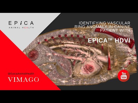
4. CT Scan: CT imaging of the chest provides the most detailed information about the specific vascular anatomy responsible for the ring. It allows the surgeon to identify precisely which vessels are involved before the procedure begins, which is particularly important for less common forms of vascular ring anomaly where the anatomy is more variable.
5. Endoscopy: Oesophagoscopy (camera examination of the oesophagus) can visualise the compression site directly and assess the degree of oesophageal dilation and any secondary mucosal changes.
6. Blood Tests: A full blood count and biochemistry panel assess the puppy’s nutritional status, organ function, and readiness for anaesthesia.
Treatment Options for Vascular Ring Anomalies in Dogs
Surgical correction is the primary and recommended treatment for vascular ring anomalies in dogs. The goal of surgery is to divide the constraining vascular structure (most commonly the ligamentum arteriosum in PRAA cases) and release the compression on the oesophagus, allowing food to pass through normally.
1. Surgical Procedure: The surgery is performed through an incision in the left side of the chest (left lateral thoracotomy) and involves identifying and ligating (tying off) the constraining band before dividing it. In PRAA, this means ligating and cutting the ligamentum arteriosum. In most cases, the procedure is technically straightforward for an experienced veterinary surgeon, though access to a soft tissue or thoracic surgery specialist is recommended for the best outcomes.
2. Timing of Surgery: Early surgical intervention, ideally before the oesophagus has been stretched and permanently damaged by prolonged dilation, produces the best outcomes. Puppies operated on at a young age before significant oesophageal dilation has established typically recover better swallowing function than those operated on after months of chronic distension.
3. Post-Surgical Feeding Management: Even after surgery successfully removes the compression, the oesophagus does not immediately return to normal diameter. The stretched, dilated segment of oesophagus above the former obstruction requires time to regain some of its normal tone, and in some dogs it never fully returns to a normal size. During recovery and beyond, special feeding strategies are important to minimise regurgitation and aspiration risk.
4. Aspiration Pneumonia Treatment: If the puppy has developed aspiration pneumonia prior to surgery, this must be assessed and treated before the surgical procedure is undertaken, as pneumonia increases anaesthetic and surgical risk. A course of antibiotics under veterinary guidance is typically required.
Puppies and dogs recovering from vascular ring surgery, and those managing ongoing oesophageal challenges, often experience stress around feeding times and unfamiliar routines. A calm, consistent feeding environment significantly supports recovery. VOSD Anxiety Care is gently formulated to support dogs experiencing stress and unsettled behaviour during recovery. Always consult your vet before introducing any supplement alongside post-surgical medications.
Related Products

Prognosis and Long-Term Outlook
The prognosis for dogs with vascular ring anomalies that receive early surgical correction is generally good to excellent for improvement in clinical signs. Most puppies show marked reduction in regurgitation frequency following surgery, begin to gain weight normally, and go on to live comfortable, active lives.
However, the degree of long-term improvement depends significantly on the extent of oesophageal dilation that had developed before surgery. Dogs operated on early, before severe, permanent oesophageal stretching has occurred, typically achieve the best functional outcomes. Dogs with significant pre-surgical oesophageal dilation may continue to have some degree of regurgitation and require lifelong feeding management, even after successful surgical release of the constriction.
The risk of aspiration pneumonia is reduced but not eliminated after surgery in dogs with residual oesophageal dilation. Careful feeding management, as described below, is the most important ongoing tool for minimising this risk.
Caring for a Dog Recovering from Vascular Ring Surgery
Consistent and careful post-surgical care makes a significant difference to functional recovery:
- Upright or elevated feeding: Feed your dog in an upright position, with the front of the body elevated at a 45 to 90-degree angle, and keep them upright for at least 10 to 15 minutes after each meal. A Bailey chair, a purpose-designed elevated feeding station, or a similar support structure helps maintain this position comfortably. Gravity assists food movement through the still-dilated oesophagus into the stomach.
- Small, frequent meals: Offering smaller amounts of food more frequently reduces the volume of food in the oesophagus at any one time, lowering the risk of regurgitation.
- Appropriate food consistency: Your vet will advise on the most appropriate food texture for your dog’s specific degree of residual oesophageal dilation. Some dogs manage best with soft or moistened food formed into small balls; others do better with liquid or slurried consistency. There is individual variation and some trial is typically needed.
- Monitor for respiratory signs: Watch for coughing, nasal discharge, or laboured breathing after meals, which may indicate aspiration. Report these to your vet promptly.
- Attend all follow-up appointments: Regular reassessment of weight gain, oesophageal function, and respiratory health allows your vet to adjust the management plan as your dog grows and develops.
Vascular Ring Anomalies in Rescued Puppies
In rescue settings, puppies with vascular ring anomalies sometimes arrive after weeks or months of unrecognised regurgitation, presenting with significant underweight, repeated respiratory infections, and in some cases established aspiration pneumonia. These puppies require stabilisation, treatment of any respiratory infection, and nutritional support before surgical assessment can be undertaken.
Early veterinary evaluation of any adopted puppy showing consistent regurgitation, poor weight gain, or coughing after meals is strongly recommended. The sooner the condition is identified, the better the surgical outcomes and the lower the risk of permanent oesophageal damage.
When Should Pet Parents Seek Veterinary Help?
Contact your veterinarian promptly if your puppy or young dog shows any of the following:
- Consistent regurgitation of undigested food after meals, without abdominal straining
- Failure to gain weight normally despite a good appetite
- Coughing or nasal discharge that develops or worsens after eating
- Visible swelling or fullness at the base of the neck after feeding
- Lethargy and weakness in a growing puppy that is not thriving at the expected rate
Vascular ring anomalies in dogs are entirely manageable with early diagnosis and appropriate surgical treatment. The earlier the condition is identified and the sooner surgery is performed, the better the chances of achieving a normal quality of life. If your puppy is showing any of the above signs, please seek veterinary assessment without delay.


