

Ventricular premature contractions in dogs, commonly abbreviated as VPCs, are extra heartbeats that arise from the ventricles (the heart’s lower pumping chambers) rather than from the sinoatrial node, the heart’s natural pacemaker. These beats occur earlier than the next expected beat in the normal rhythm, creating a characteristic irregular pulse pattern. VPCs are among the most commonly detected cardiac rhythm abnormalities in dogs, and their significance varies enormously depending on how frequently they occur, whether they appear in isolation or in runs, and what underlying condition may be driving them. For many dogs, occasional VPCs require nothing more than monitoring. For others, they are an important warning sign of significant underlying heart disease. Understanding the difference is what determines the right response.
What Are Ventricular Premature Contractions in Dogs?
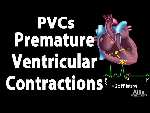
In a normal heartbeat, the electrical impulse that triggers ventricular contraction travels down from the sinoatrial node, through the atrioventricular node, and then through the bundle branches to activate both ventricles simultaneously in a precisely timed sequence. A ventricular premature contraction occurs when an irritable focus of tissue within the ventricular myocardium generates its own electrical impulse ahead of schedule, triggering an extra heartbeat that interrupts the normal rhythm.
Because this extra beat originates from an abnormal location in the ventricles rather than from the normal conduction pathway, it produces a wider and differently shaped QRS complex on the electrocardiogram (ECG) than a normal beat. This distinctive ECG appearance is what allows veterinarians to identify and count VPCs during cardiac monitoring.
After a VPC, there is typically a compensatory pause before the next normal beat resumes, because the normal sinus impulse that would have occurred at that moment arrives while the ventricles are still in their refractory (unresponsive) period following the premature beat. It is this pause that pet parents sometimes describe as a “skipped beat” sensation when they feel their dog’s pulse.
How VPCs Affect the Heart
The clinical impact of VPCs depends on their frequency and pattern:
- Occasional, isolated VPCs: Single VPCs occurring infrequently in an otherwise healthy heart often cause no significant reduction in cardiac output and produce no clinical signs. They may be entirely benign.
- Frequent VPCs: When VPCs occur many times per hour or make up a significant proportion of the total heartbeats over a 24-hour period, they can begin to reduce cardiac efficiency and may produce symptoms such as weakness or exercise intolerance.
- Runs of VPCs (ventricular tachycardia): When three or more VPCs occur consecutively, this constitutes a run of ventricular tachycardia. If runs are frequent, prolonged, or very rapid, they can cause significant reduction in cardiac output and may degenerate into ventricular fibrillation in severe cases.
- R-on-T phenomenon: When a VPC falls on the T wave of the preceding normal beat, it can trigger ventricular fibrillation. This is a recognised high-risk pattern on ECG that warrants prompt treatment.
Symptoms of Ventricular Premature Contractions in Dogs
The symptoms of VPCs in dogs vary significantly based on their frequency and the presence of any underlying cardiac disease:
- Many dogs with occasional or even moderately frequent VPCs show no visible symptoms at all. The abnormal rhythm is detected only during a routine veterinary examination when the vet notices an irregular pulse or characteristic sounds through the stethoscope.
- Weakness or mild lethargy, particularly in dogs with frequent VPCs reducing cardiac efficiency
- Exercise intolerance, tiring more quickly than expected
- Fainting (syncope) or sudden collapse in dogs with frequent runs of ventricular tachycardia causing significant drops in cardiac output
- An irregular pulse detected when you feel your dog’s heartbeat on the chest or pulse in the groin
- Restlessness or apparent awareness of palpitations in some dogs
Irregular heartbeats are one of the more common findings picked up during routine annual health examinations in older dogs, often before any clinical symptoms have developed. This is one reason why regular veterinary check-ups are particularly valuable for middle-aged and senior dogs, particularly those of breeds predisposed to cardiac conditions.
Causes of Ventricular Premature Contractions in Dogs
VPCs in dogs can arise from a wide range of underlying causes, and identifying the specific trigger is an important part of the diagnostic workup:
- Primary cardiac disease: Cardiomyopathy is one of the most important underlying causes of VPCs in dogs. Arrhythmogenic right ventricular cardiomyopathy in Boxers and Bulldogs, dilated cardiomyopathy in large breeds including Dobermanns, Great Danes, and Irish Wolfhounds, and advanced mitral valve disease with significant left ventricular remodelling can all produce ventricular ectopic activity.
- Splenic disease: Splenic masses, particularly splenic haemangiosarcoma, are a well-recognised cause of VPCs in dogs. The mechanism is thought to involve myocardial contusion from abdominal haemorrhage and autonomic activation. Any dog presenting with VPCs should have an abdominal assessment to rule out splenic abnormalities.
- Electrolyte imbalances: Abnormal potassium, calcium, or magnesium levels destabilise the electrical threshold of ventricular myocytes and increase the likelihood of ectopic firing. Hypokalaemia (low potassium) is a particularly common cause in dogs on diuretics for cardiac or other conditions.
- Tick-borne infections: Ehrlichia, Babesia, and other tick-borne diseases cause significant systemic inflammation, myocardial involvement, and electrolyte disturbances that can drive VPCs. In India, where tick-borne disease is prevalent in urban and peri-urban dog populations, this is a clinically important cause that is often overlooked.
- Gastric dilatation and volvulus (GDV): VPCs are extremely common in dogs with GDV and may persist for 24 to 72 hours after surgical correction. Monitoring and anti-arrhythmic treatment are part of standard GDV post-operative care.
- Trauma: Myocardial contusion from road traffic accidents or significant blunt chest trauma can cause VPCs that typically appear 24 to 72 hours after the injury, sometimes when the dog appears otherwise stable.
- Drug reactions and toxins: Certain medications, including digoxin at excessive doses, and some toxins, can sensitise the ventricular myocardium to ectopic firing.
- Idiopathic VPCs: In some dogs, particularly German Shepherds with inherited ventricular arrhythmia, VPCs occur without any identifiable underlying cause. German Shepherd juvenile ventricular arrhythmia is a recognised breed-specific condition.
Related Videos


How Vets Diagnose Ventricular Premature Contractions in Dogs
Diagnosis begins at the physical examination and is confirmed with targeted cardiac testing:
1. Physical Examination and Auscultation: An irregular pulse, premature beats, or a characteristic sound pattern on auscultation may suggest VPCs. The vet will also assess for abdominal distension (splenic disease), fever (infection), and any other clinical signs that point toward an underlying cause.
2. Electrocardiogram (ECG): The ECG confirms the presence of VPCs by demonstrating wide, bizarre QRS complexes that occur earlier than expected and are not preceded by a normal P wave. The ECG also assesses whether VPCs are isolated or occur in runs, and identifies any concerning patterns such as R-on-T or multiform VPCs (which originate from multiple ectopic foci and indicate a more irritable myocardium).
3. Holter Monitor: Because VPCs can be intermittent, a 24-hour ambulatory Holter monitor is the most comprehensive tool for counting VPC frequency, characterising their pattern, and determining whether runs of ventricular tachycardia are occurring. Published veterinary guidelines use Holter-derived VPC counts to guide treatment decisions in predisposed breeds such as Dobermanns and Boxers.
4. Echocardiography: An echocardiogram assesses cardiac structure and function, identifying underlying cardiomyopathy, structural abnormalities, or changes in ventricular function that may be driving the arrhythmia.
5. Abdominal Ultrasound: Recommended in all dogs with newly identified VPCs to assess the spleen and other abdominal organs for masses or abnormalities.
6. Blood Tests: Electrolyte levels, complete blood count, biochemistry panel, and tick-borne disease screening (particularly Ehrlichia and Babesia serology) are all part of a thorough workup.
Treatment for Ventricular Premature Contractions in Dogs
Treatment decisions for VPCs in dogs are guided by the frequency and pattern of arrhythmia, the presence of clinical signs, and the nature of the underlying cause:
| VPC Pattern | Clinical Signs | Typical Management Approach |
|---|---|---|
| Occasional isolated VPCs, no underlying disease | None | Monitor with periodic Holter rechecks; no medication typically required |
| Frequent VPCs, no clinical signs, underlying disease identified | None or mild | Treat underlying cause; reassess VPC frequency after treatment |
| Frequent VPCs with clinical signs (weakness, exercise intolerance) | Present | Anti-arrhythmic medication (sotalol, mexiletine) plus treatment of underlying cause |
| Runs of ventricular tachycardia, with or without signs | Variable | Anti-arrhythmic medication indicated; urgent if R-on-T or haemodynamically unstable |
| Post-GDV or post-trauma VPCs | Variable | Continuous ECG monitoring; anti-arrhythmic treatment if rapid runs develop |
Anti-arrhythmic medications commonly used in dogs with significant VPCs include sotalol, mexiletine, and in some cases lidocaine (for acute intravenous management). The choice of drug depends on the arrhythmia characteristics, the underlying cause, and the dog’s overall health. Medication selection and dosing must be guided by a veterinarian or veterinary cardiologist.
Treatment of the underlying cause is always the most important step. VPCs from tick-borne disease resolve when the infection is treated. VPCs from hypokalaemia resolve when potassium is corrected. VPCs from a splenic mass may require surgical removal of the spleen.
In dogs where stress contributes to VPC frequency, particularly rescued dogs or those in high-stimulation environments, a calmer home routine supports overall cardiac stability. VOSD Anxiety Care is gently formulated to support dogs experiencing stress and unsettled behaviour. Always consult your vet before introducing any supplement alongside anti-arrhythmic medications.
For dogs in India where tick-borne infections are a significant cause of arrhythmias, consistent tick prevention is one of the most important preventive steps available. VOSD Spot-On Tick and Flea Protection with IGR provides reliable protection against ticks and the infections they carry. Always use as directed and discuss with your vet.
Related Products


Prognosis for Dogs with Ventricular Premature Contractions
The prognosis for VPCs in dogs depends primarily on the underlying cause and the frequency and pattern of the arrhythmia:
- Dogs with occasional, isolated VPCs and no identifiable underlying disease typically have an excellent prognosis. Many of these dogs never develop significant arrhythmia-related symptoms and can be monitored safely over the long term.
- Dogs whose VPCs are caused by a reversible condition, such as tick-borne infection, electrolyte disturbance, or a resectable splenic mass, generally achieve good resolution of the arrhythmia once the primary condition is treated.
- Dogs with VPCs secondary to progressive cardiomyopathy have a prognosis determined primarily by the nature and stage of the underlying cardiac disease. Anti-arrhythmic therapy can reduce the risk of sudden cardiac death and improve quality of life, but cannot reverse the underlying muscle disease.
- Dogs with German Shepherd juvenile ventricular arrhythmia most commonly experience spontaneous improvement or resolution of the arrhythmia as they pass through adolescence, though sudden death can occur in severely affected individuals during the highest-risk period.
Living with a Dog with VPCs
- Attend all Holter monitoring appointments: Periodic Holter rechecks track whether VPC frequency is increasing, stable, or improving, and guide the timing of medication introduction or adjustment.
- Avoid excessive exertion if symptomatic: Dogs with frequent VPCs or runs of ventricular tachycardia should have strenuous exercise restricted until the arrhythmia is controlled. Discuss appropriate activity levels with your vet.
- Give all medications consistently: Anti-arrhythmic drugs must be given at the same time each day without interruption. Inconsistent dosing can allow breakthrough arrhythmias.
- Keep a fainting diary: Note any episodes of weakness, collapse, or fainting with timing and duration. This is valuable information for your vet at follow-up appointments.
- Tick prevention year-round: In areas where tick-borne disease is present, consistent tick control is one of the most practical protective measures you can take for your dog’s cardiac health.
When to See a Vet
Contact your veterinarian promptly if your dog shows any of the following:
- Any episode of fainting or sudden collapse
- Weakness, exercise intolerance, or unusual lethargy that has appeared or worsened recently
- An irregular pulse that you can detect yourself when feeling the heartbeat
- Known cardiac disease with new or worsening symptoms
- Any systemic signs of illness alongside an irregular heartbeat, including fever, reduced appetite, or lethargy
Ventricular premature contractions in dogs span a wide spectrum from benign to clinically significant. The right response depends entirely on accurate diagnosis, careful assessment of frequency and pattern, and thorough investigation of the underlying cause. Most dogs with well-characterised and appropriately managed VPCs continue to live comfortable, active lives. Early detection through regular veterinary examinations remains the most reliable way to ensure that any change in VPC burden is identified promptly.


