

Skin tumors are among the most frequently diagnosed growths in dogs. Some are harmless. Some are not. And the only way to know which you are dealing with is to have them properly examined.
Basal cell tumors are one of the more common skin growths found in dogs, and the good news is that the vast majority of them are benign. They grow slowly, they tend to stay localised, and when treated appropriately, most dogs recover completely.
But benign does not mean ignorable. Left unattended, even a non-malignant tumor can grow large enough to ulcerate, become infected, and cause real discomfort. And without a proper diagnosis, there is no way to distinguish a benign basal cell tumor from a malignant one on appearance alone.
This guide explains what basal cell tumors are, how they present, and what veterinary care looks like from diagnosis through recovery. If you have noticed a new lump on your dog’s skin, understanding lumps, bumps, and cysts on dogs is a useful first step before diving deeper into any specific tumor type.
What Are Basal Cell Tumors in Dogs?
To understand where these tumors come from, it helps to understand the structure of the skin.
The skin is composed of multiple layers. The outermost layer, the epidermis, has a deepest stratum called the basal layer. This layer contains basal cells, which are responsible for continuously generating new skin cells that migrate upward, mature, and eventually shed from the surface. It is a process of constant renewal that keeps the skin intact and functional.
When basal cells begin dividing abnormally, they can form a tumor. In most cases, this tumor is benign, meaning the cells are not invasive and the growth does not spread to other parts of the body. These are true basal cell tumors.
In a small number of cases, the cells become malignant. This form is called basal cell carcinoma, and it behaves very differently from its benign counterpart. It can invade surrounding tissue and, in rare cases, spread to other organs.
Basal cell tumors most commonly appear as solitary, raised, firm lumps on the head, neck, or shoulders, though they can develop anywhere on the body. They are more frequently diagnosed in middle-aged to older dogs and tend to be well-defined and slow-growing. Understanding skin cancer in dogs more broadly helps contextualise where basal cell tumors sit within the wider spectrum of canine skin conditions.
Symptoms of Basal Cell Tumors in Dogs
The appearance of basal cell tumors can vary, which is part of why veterinary confirmation matters.
A dome-shaped or raised lump on the skin. This is the most typical presentation. The lump is usually firm to the touch and clearly defined from surrounding tissue.
Hairless surface over the mass. The skin overlying the tumor often loses its fur, leaving a smooth or slightly shiny surface.
Dark or pigmented appearance. Many basal cell tumors are darkly pigmented, appearing brown, grey, or black. This pigmentation can sometimes make them appear more alarming than they are, but colour alone does not confirm malignancy.
Ulceration or bleeding. If the tumor grows large enough or is repeatedly traumatized by the dog scratching or rubbing the area, the surface can break down. Open sores that do not heal should always be examined promptly.
Discharge from the lesion. Ulcerated tumors may produce a clear or slightly bloody discharge.
Size variation. These tumors can range from small, pea-sized nodules to significantly larger masses several centimetres across. Size at the time of discovery depends largely on how long the tumor has been growing and how attentive the owner has been to skin changes.
Irritation or self-trauma. Dogs may lick, scratch, or rub at the area if the tumor causes itching or discomfort, which in turn worsens the skin condition around it.
For any new skin growth showing these characteristics, a veterinary assessment is the appropriate response. Detailed guidance on identifying various types of skin changes is available in the ” Lumps, bumps, and Cysts on Dogs ” resource.
Causes and Risk Factors
The precise cause of basal cell tumor development in dogs is not fully established, but several contributing factors have been identified.
Genetics appears to play a meaningful role. Certain breeds show a notably higher incidence of basal cell tumors compared to the general dog population. Siberian Huskies, Shetland Sheepdogs, Cocker Spaniels, and several terrier breeds are among those with a higher predisposition. This pattern suggests an inherited vulnerability in the basal cell layer of the skin.
Age is a consistent factor. These tumors are far more commonly diagnosed in middle-aged and older dogs. The cumulative effect of cellular wear over time increases the likelihood of abnormal cell division.
Sun exposure has been theorised as a contributing environmental factor, particularly for tumors appearing on sparsely haired or lightly pigmented areas. Dogs that spend significant time outdoors with exposure to ultraviolet radiation may have a somewhat higher risk, though this is less definitively established in dogs than it is in humans.
Hormonal influences and chronic skin irritation have been considered as potential contributing factors in some cases, though neither has been conclusively proven as a direct cause.
What is clear is that no single owner action reliably prevents these tumors from forming. The most effective response to the risk factors is regular skin monitoring so that any new growth is identified and assessed early.
Related Videos


How Veterinarians Diagnose Basal Cell Tumors
A lump that looks like a basal cell tumor cannot be confirmed as one by visual inspection alone. The cells must be examined to know what you are dealing with.
Physical examination. The veterinarian will assess the lump’s size, location, texture, surface characteristics, and any changes in the surrounding skin. They will also check nearby lymph nodes for any signs of swelling that might suggest malignancy.
Fine needle aspiration (FNA). This is typically the first diagnostic step. A thin needle is inserted into the mass to collect a small sample of cells, which are then examined under a microscope. FNA is quick, requires minimal sedation, and can provide useful initial information. For many basal cell tumors, cytology from FNA is informative, though a definitive diagnosis often requires more tissue.
Biopsy and histopathology. A surgical biopsy, where a tissue sample is sent to a pathology laboratory, provides the definitive diagnosis. The pathologist examines the cellular architecture and can confirm whether the tumor is benign or malignant, and how its cells are structured. This information directly shapes treatment decisions.
Imaging. For tumors suspected of being malignant, chest X-rays and abdominal ultrasound help assess whether spread has occurred. This is not typically necessary for straightforward benign cases but becomes important if malignancy is suspected from the biopsy.
The diagnostic process is thorough for good reason. A definitive answer changes the entire conversation about treatment and prognosis.
Treatment Options for Basal Cell Tumors
Once the tumor has been assessed and confirmed, treatment planning is usually straightforward for benign cases.
Surgical removal is the primary treatment for basal cell tumors and is typically curative for benign forms. The goal is to excise the tumor with clear margins, ensuring no abnormal cells remain at the edges of the removed tissue. For most benign basal cell tumors, a single surgical procedure achieves complete removal and the dog does not require further cancer-specific treatment.
Cryosurgery is an alternative for small, superficial tumors that are clearly benign. This technique uses extreme cold to destroy the abnormal cells without conventional surgery. It is less commonly used than excision but may be appropriate in selected cases, particularly in older dogs where general anaesthesia carries higher risk.
Active monitoring may occasionally be recommended for very small, clearly benign tumors in elderly dogs where the risks of surgery outweigh the risks of leaving the growth in place. This approach requires strict discipline around monitoring for any changes in size, shape, or surface integrity.
Additional therapies for malignant tumors. When histopathology reveals basal cell carcinoma rather than a benign tumor, the approach changes significantly. Surgery remains central but must achieve wider margins. Radiation therapy may be used when complete excision is not possible. Chemotherapy has a more limited role in basal cell carcinoma compared to other canine skin cancers, but may be considered in advanced cases. Consulting a veterinary oncologist is strongly recommended for any confirmed malignant diagnosis.
Related Products


Recovery After Tumor Removal
For dogs that have undergone surgical removal of a benign basal cell tumor, recovery is generally uncomplicated.
Most dogs go home the same day or the day after surgery. The surgical site will require monitoring for the first one to two weeks. Owners should check daily for signs of infection such as redness, swelling, discharge, or heat around the wound.
An Elizabethan collar, often called a cone, is typically recommended to prevent the dog from licking or chewing at the surgical site. This is one of the most important post-operative instructions and one of the most frequently ignored. Dogs licking their wounds is not helpful and can introduce infection or disrupt the healing tissue.
Activity restriction during the recovery period prevents excessive movement from disrupting the wound closure. Short, calm walks on a lead are generally acceptable. Rough play, jumping, and swimming should be avoided until the wound has fully healed.
If antibiotics are prescribed, the full course must be completed even if the wound appears to be healing well.
A follow-up appointment is usually scheduled to check healing and discuss histopathology results if not already received.
Prognosis for Dogs With Basal Cell Tumors
For the vast majority of dogs with benign basal cell tumors, the prognosis following surgical removal is excellent.
Complete excision with clean margins is typically curative. Recurrence after complete removal is uncommon. Most dogs go on to live normal, healthy lives with no further issues related to the tumor.
The small percentage of cases involving basal cell carcinoma carry a more guarded prognosis, particularly if the cancer has spread regionally or is not amenable to complete surgical removal. However, even malignant basal cell tumors are considered less aggressive than many other canine skin cancers, and meaningful survival times are achievable with appropriate treatment.
The most important determinant of outcome, in both benign and malignant cases, is how early the tumor is identified and treated. A small, well-defined tumor caught early is almost always a far simpler clinical problem than one that has grown large, ulcerated, or begun to invade surrounding tissue.
Possible Complications if Untreated
Choosing to ignore a skin tumor, even one that appears benign, carries real risks over time.
Ulceration. As the tumor grows, the skin over it can break down, forming open sores that are painful and vulnerable to infection.
Secondary infection. Open ulcerated tumors are an entry point for bacteria. Infected tumors require treatment with antibiotics and significantly complicate any subsequent surgical management.
Tissue damage. Larger tumors can involve deeper tissue layers, making surgical removal more complex and increasing the likelihood of complications during excision.
Malignant transformation. While uncommon, benign tumors can theoretically undergo malignant change over time. Regular monitoring and timely removal remove this possibility entirely.
Chronic discomfort. A growing mass in a sensitive location causes ongoing irritation. Dogs that repeatedly scratch, lick, or rub at a tumor suffer unnecessary discomfort that could be avoided with early treatment.
Other Skin Tumors That Look Similar
Basal cell tumors share visual characteristics with several other skin growths, which is precisely why professional diagnosis matters.
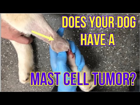
Mast cell tumors are one of the most common and most dangerous skin tumors in dogs. They can appear as raised, firm skin lumps that closely resemble benign basal cell tumors. However, mast cell tumors are malignant and require a very different treatment approach. They cannot be reliably distinguished from basal cell tumors by appearance alone.
Sebaceous cysts are benign, fluid-filled sacs that form when a hair follicle or sebaceous gland becomes blocked. They are soft rather than firm and generally harmless, but can become infected or rupture.
Histiocytomas are benign tumors most commonly seen in young dogs. They appear suddenly, grow quickly, and often resolve on their own within a few months. They can alarm owners due to their rapid appearance but are generally not a cause for concern.
Lipomas are benign fatty tumors found beneath the skin. They feel soft and mobile rather than firm and fixed.
The consistent message across all of these is simple: do not guess. A veterinary diagnosis is the only reliable way to know what a lump is and what it requires.
When Should You See a Veterinarian?
Any new lump on your dog’s skin warrants a veterinary examination. The threshold for booking an appointment should be low.
See your vet promptly if you notice a lump that was not there before, a growth that has changed in size, shape, or surface texture over a short period, any skin mass that has become ulcerated, is bleeding, or is producing discharge, a lump that your dog is repeatedly attending to through licking or scratching, or any skin change accompanied by lethargy, weight loss, or reduced appetite.
Early assessment does not necessarily mean aggressive treatment. For many benign basal cell tumors, the veterinarian may recommend removal, monitoring, or simply confirm what the growth is and advise accordingly. What early assessment always provides is clarity, and clarity allows you to make the right decision for your dog.
Early Detection Is Key
Basal cell tumors in dogs are, in most cases, a manageable and treatable condition. The overwhelming majority are benign. Surgical removal is typically curative. Recovery is straightforward. And with early intervention, most dogs are entirely unaffected by them in the long run.
But that outcome depends on one thing: finding the tumor while it is still small, still localised, and still easily addressed.
Regular skin checks at home, combined with routine veterinary examinations, are the most reliable way to stay ahead of any skin changes your dog may develop. Run your hands across your dog’s body regularly. Pay attention to new lumps, changes in existing ones, and any areas your dog seems to be attending to more than usual.
If you find something new, do not wait and watch indefinitely. Have it examined.
For a complete overview of how to identify and interpret different types of skin growths in dogs, skin cancer in dogs and the VOSD veterinary resource library provide detailed, clinically grounded guidance to help you make informed decisions about your dog’s health.


