

If you see it, you cannot ignore it.
A red, tube-like mass of tissue protruding from your dog’s anus is one of the most visually alarming things a pet parent can encounter. It is also one of the clearest medical emergencies in veterinary practice. Rectal prolapse is not a condition to photograph and research at home. It is a condition that requires veterinary care within hours, because the exposed tissue begins to dry out, swell, and lose its blood supply the moment it is outside the body.
Every minute of delay increases the likelihood of permanent tissue damage, more complex treatment, and a significantly harder recovery.
What Is Rectal Prolapse in Dogs?
Rectal prolapse occurs when one or more layers of the rectal wall are forced through the anal opening and remain visible outside the body. The rectum, which is the final segment of the large intestine, is pushed outward by straining pressure until it protrudes beyond the anus.
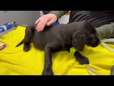
It presents as a cylindrical, red or pink, moist tube of tissue that may appear during or after straining and either retreats spontaneously or remains visible even at rest.
Incomplete Prolapse
In incomplete prolapse, only the inner mucosal lining of the rectum protrudes, typically visible during straining and potentially retracting when the straining episode resolves. This form may be intermittent and is sometimes mistaken for haemorrhoids, which do not occur in dogs.
Complete Prolapse
In complete prolapse, the full thickness of the rectal wall protrudes and remains visible at rest. The tissue is a distinct cylindrical mass with a central opening, which is the continuation of the rectal lumen. This presentation is always an emergency.
Symptoms of Rectal Prolapse in Dogs
- A red, pink, or increasingly darkened cylindrical mass protruding from the anus
- Straining and repeated unsuccessful attempts to defecate
- Obvious pain or discomfort, particularly in the perianal region
- Persistent licking or biting at the rear end
- Bleeding or mucoid discharge from the protruding tissue
- Restlessness and inability to settle comfortably
- In advanced cases, lethargy and systemic signs of deterioration
The colour of the tissue is one of the most important clinical indicators. Fresh, pink or bright red tissue that is still receiving adequate blood supply is far more amenable to manual reduction and preservation than dark red, purple, or black tissue, which indicates compromised circulation and progressive necrosis.
Causes of Rectal Prolapse in Dogs
The common denominator in virtually all rectal prolapse cases is straining. Sustained, forceful straining raises the intra-abdominal and intrarectal pressure to a point where the rectal wall is pushed outward through the anal opening.
Primary causes include severe or persistent diarrhoea, which produces repeated straining without adequate stool bulk to support normal defecation mechanics, chronic constipation requiring prolonged effort to pass impacted faecal material, and intestinal parasites. Parasitic infections, particularly whipworm and roundworm, are a leading cause in puppies, where the combination of heavy parasitic burden, soft stools, and the underdeveloped anal sphincter creates significant prolapse risk.
Secondary causes include diseases of the lower intestinal tract that produce chronic straining, urinary obstruction causing effort that translates into rectal pressure, difficult or prolonged labour in female dogs, rectal tumours and polyps that stimulate persistent straining reflexes, and perineal hernia altering normal defecation mechanics.
In every case, identifying and treating the underlying cause is as important as managing the prolapse itself. Reduction of the prolapse without addressing the straining trigger produces reliable recurrence.
Related Videos


How Veterinarians Diagnose Rectal Prolapse in Dogs
The prolapse itself is diagnosed visually. The cylindrical mass with a palpable central lumen, distinguishing it from other perianal masses that do not have an open centre, confirms rectal prolapse on physical examination.
The more important diagnostic work focuses on establishing why the prolapse occurred. This requires a thorough rectal examination to assess the extent and viability of the prolapsed tissue and the condition of the anal sphincter, faecal testing to identify parasites as a cause, particularly in young dogs, abdominal imaging through ultrasound and radiography to assess the intestinal tract for masses, obstruction, or disease, and blood tests to evaluate for systemic disease contributing to chronic straining.
Understanding the broader category of lower intestinal disorders, including abnormal growths in the lower intestines of dogs, is clinically relevant because these conditions are among the common drivers of the chronic straining that precipitates prolapse.
| Stage | Clinical Presentation | Required Action |
|---|---|---|
| Early | Mild protrusion visible only during straining, tissue pink and moist | Immediate veterinary assessment, manual reduction attempted |
| Moderate | Persistent visible prolapse at rest, tissue swollen but viable | Medical management, manual reduction under anaesthesia, purse-string suture |
| Severe | Significant swelling, bleeding, pain, tissue beginning to darken | Emergency treatment, reduction attempted if tissue viable, surgical preparation |
| Critical | Tissue dark purple or black, signs of necrosis, systemic deterioration | Emergency surgery, resection of non-viable tissue, intensive supportive care |
Treatment for Rectal Prolapse in Dogs
Immediate Care
The moment a prolapse is identified, the priority is protecting the exposed tissue from further damage during transport to the veterinary clinic. Keeping the tissue moist with a clean, damp cloth or gentle application of a water-based lubricant reduces drying and surface trauma. Do not attempt forceful manual reduction at home.
Medical Treatment (Manual Reduction)
When the prolapsed tissue is still viable, meaning it is pink or red, moist, and not showing signs of necrosis, manual reduction under general anaesthesia is the first-line treatment. The tissue is cleaned, lubricated, and gently repositioned back through the anal opening.
Following successful reduction, a purse-string suture is placed around the anal opening, tightening it sufficiently to retain the reduced rectum while still allowing passage of soft stools. The suture is maintained for several days while swelling resolves and the rectal tissue re-establishes its normal position.
Medical management of swelling and pain follows reduction, alongside specific treatment of the underlying cause. Without addressing the cause, recurrence after reduction is the expected outcome rather than the exception.
Surgical Treatment
When the prolapsed tissue is no longer viable, when manual reduction fails, or when the prolapse has recurred multiple times, surgery is required. Surgical options include resection of the non-viable segment of prolapsed rectum, followed by anastomosis of the healthy rectal ends, and colopexy, a procedure that anchors the descending colon to the abdominal wall to prevent further prolapse.
Colopexy is particularly recommended in recurrent cases and in dogs where the underlying cause cannot be fully resolved, as it provides mechanical support that reduces the likelihood of re-prolapse regardless of straining episodes.
Related Products


Prognosis
The prognosis for rectal prolapse is good when treatment is provided promptly and the tissue is still viable at the time of intervention. Dogs treated within a few hours of prolapse with successful manual reduction, appropriate suturing, and management of the underlying cause typically recover fully with a low recurrence rate.
The prognosis worsens significantly with delay. Tissue that has lost its blood supply cannot be reduced and requires surgical resection, which carries greater operative risk, a longer recovery period, and the potential for complications, including stricture formation at the surgical site.
Recurrence is the most common long-term challenge, particularly when the straining cause is chronic or not fully resolvable. In these cases, colopexy at the time of surgical management significantly reduces the recurrence rate.
Why Rectal Prolapse Is an Emergency
This is a point that cannot be overstated. The exposed rectal tissue begins to sustain damage the moment it is outside the body.
Without the protection of the anal canal and the moisture of the rectal environment, the tissue dries and swells progressively. Swelling impairs venous return, which worsens congestion, which accelerates further swelling. The blood supply to the prolapsed segment becomes compromised, and tissue death begins in the most distal, most exposed portions first.
Once tissue is necrotic, it cannot be reduced. It must be surgically removed. And beyond the local tissue damage, the bacterial translocation from the devitalised, exposed rectal mucosa creates systemic infection risk.
The difference between a successful manual reduction and a major surgical resection is frequently measured in hours.
When to See a Veterinarian
Seek emergency veterinary care immediately if your dog shows any of the following:
- Any visible mass or tissue protruding from the anus, regardless of size or apparent severity
- A dog that is straining intensely and producing no stool, particularly alongside visible perianal swelling
- Bleeding from the anus accompanied by straining
- The prolapsed tissue changing colour from pink to dark red, purple, or black
- Lethargy, weakness, or signs of systemic illness alongside any perianal abnormality
There is no safe window for observation at home when prolapsed tissue is visible. This is a veterinary emergency from the moment you identify it.
Prevention and Risk Reduction
The most consistent preventive strategy is eliminating the straining that causes prolapse.
Treat diarrhoea early and do not allow persistent loose stools to continue without investigation. Loose, frequent stools produce repeated defecation attempts and repeated straining that progressively stress the anal sphincter and rectal support structures.
Regular deworming is essential, particularly in puppies and young dogs, where parasitic infections are a primary cause of the chronic straining that leads to prolapse. Maintaining a current, veterinarian-recommended parasite control protocol directly reduces this risk category.
Manage chronic constipation through appropriate dietary fibre content, adequate hydration, and prompt veterinary investigation of any dog that is consistently straining to defecate. Do not allow prolonged constipation to continue without professional assessment.
Monitor your dog’s defecation behaviour as part of normal daily observation. A dog that is straining, taking longer than usual to defecate, or repeatedly attempting without success is communicating that something is wrong. Early investigation of these signs consistently prevents the escalation that leads to prolapse. For dogs with concurrent lower intestinal disease, addressing conditions such as rectoanal polyps in dogs and intestinal leiomyoma in dogs that drive straining is directly preventive.


