When a dog strains to pass stool, the immediate assumption is constipation. But straining is a symptom, not a diagnosis. And in some dogs, what looks like a routine digestive issue is caused by an abnormal growth inside the rectum that is physically obstructing normal defecation.
Rectoanal polyps are abnormal tissue growths that arise from the mucosal lining of the rectum or the anal canal. They are most often benign, meaning they do not spread to other organs, but benign does not mean harmless. A polyp that bleeds, causes pain, partially obstructs the passage of stool, or grows large enough to protrude from the anus is causing real, ongoing discomfort and carries risks that escalate with every week it goes without assessment.
Early evaluation changes the outcome significantly.
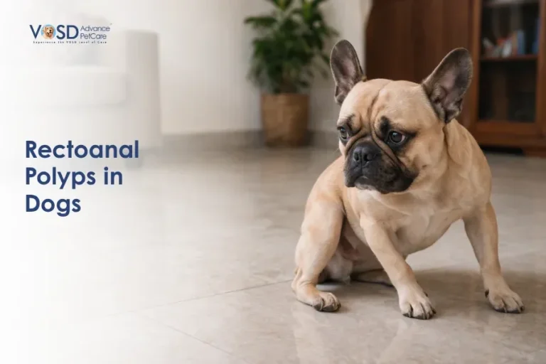
What Are Rectoanal Polyps in Dogs?
Rectoanal polyps are localised, abnormal overgrowths of the mucosal lining of the rectum or anal canal. They project into the rectal lumen as pedunculated growths attached by a stalk, or as sessile masses sitting flush against the mucosal surface.
Most polyps in dogs are benign adenomatous growths or inflammatory polyps. They do not invade surrounding tissue and do not spread to lymph nodes or distant organs. However, some polyps, particularly larger ones or those that have been present for an extended period without treatment, can undergo malignant transformation. The distinction between benign and malignant can only be made through biopsy, which is why every rectal mass requires histopathological assessment regardless of its apparent clinical behaviour.
Symptoms of Rectoanal Polyps in Dogs
The symptoms are directly produced by the presence of the growth within the rectal lumen and its effects on normal defecation.
Straining to Defecate (Tenesmus)
This is the most consistent and most commonly presenting sign. A dog that repeatedly squats, strains, and produces little or nothing, or produces small amounts with obvious effort, is experiencing tenesmus. When a polyp is the cause, the physical presence of the growth within the rectal lumen creates a sensation of incomplete evacuation and mechanical interference with normal stool passage.
This presentation is frequently misidentified as constipation, and dogs are sometimes managed with dietary changes or laxatives for extended periods before the underlying structural cause is investigated.
Blood in Stool (Hematochezia)
Polyps bleed readily because their mucosal surface is fragile and directly exposed to the mechanical trauma of passing stool. Fresh red blood in or coating the stool, sometimes appearing as streaks on the surface or as a small volume of blood expelled separately, is a consistent sign. Hematochezia alongside straining should always prompt professional rectal examination rather than home management.
Visible Mass from the Anus
Larger polyps, particularly pedunculated ones on longer stalks, may protrude through the anal opening during or after defecation. This intermittent protrusion can be mistaken for rectal prolapse, and the distinction requires veterinary examination. A mass that appears and then retreats is a characteristic feature of a pedunculated polyp being passed partially through the anal canal with straining and then returning to position.
Difficulty Passing Stool
As a polyp enlarges, it occupies progressively more of the rectal lumen. Stool passage requires navigating around the growth, which creates partial obstruction, discomfort, and altered stool character. Dogs may pass narrower stools than normal, indicating that the rectal lumen is being partially compressed.
Diarrhoea and Mucus
Rectal mucosal irritation from the presence and movement of the polyp stimulates excess mucus production and may produce loose, mucus-coated stools. This sign is often attributed to colitis before the underlying rectal growth is identified.
Pain and Discomfort During Defecation
The combination of mucosal irritation, bleeding, and partial obstruction makes defecation painful. Dogs may hesitate before attempting to defecate, vocalise during the attempt, or begin scooting and licking at the perianal area as a response to the localised discomfort.
Causes of Rectoanal Polyps in Dogs
Chronic Inflammation
Chronic inflammatory conditions of the rectal mucosa are the most consistently associated factor in canine rectal polyp development. Persistent mucosal irritation from inflammatory bowel disease, chronic colitis, or recurring rectal infection stimulates abnormal mucosal cell proliferation over time. The repeated cycles of injury and repair create the conditions for abnormal tissue overgrowth.
Genetic and Age-Related Factors
Rectoanal polyps are seen more frequently in middle-aged to older dogs, consistent with the accumulated cellular changes that increase abnormal proliferation risk with age. A genetic component is suspected in some cases, with certain breeds appearing to be represented more frequently in rectal polyp diagnoses, though this has not been definitively characterised across the canine population.
Abnormal Tissue Growth
In some cases, polyp development reflects a primary abnormality in the regulation of mucosal cell growth without an identifiable inflammatory trigger. Adenomatous polyps, arising from glandular mucosal cells, develop through this mechanism and are analogous to the adenomatous polyps seen in human colorectal disease.
Possible Malignant Transformation
While most rectoanal polyps in dogs are benign, the risk of malignant transformation increases with polyp size and duration. Larger polyps, those with irregular surfaces, and those that have been monitored without treatment over extended periods carry a higher probability of containing or developing into carcinoma. This is the most clinically significant reason why a wait-and-see approach to rectal growths is not appropriate.
Related Videos


How Rectoanal Polyps Develop
Polyp formation begins with abnormal proliferation of the rectal mucosal cells. Whether driven by chronic inflammation, genetic factors, or primary growth dysregulation, the mucosal cells multiply beyond their normal boundaries and form an outgrowth into the rectal lumen.
As the polyp grows, it projects progressively further into the lumen, occupying space that was previously available for stool passage. The blood supply within the polyp is fragile, producing bleeding with the mechanical trauma of passing stool. The polyp’s surface produces mucus, contributing to the mucoid stool changes associated with the condition. In pedunculated polyps, the stalk allows movement of the growth with rectal contractions, which is what produces the intermittent protrusion through the anal opening.
How Veterinarians Diagnose Rectoanal Polyps in Dogs
Physical and Rectal Examination
Many rectal polyps are within reach of a gloved digital rectal examination. The veterinarian can often palpate the growth directly, assessing its location, approximate size, consistency, and attachment. This examination is performed under sedation in most cases to allow thorough assessment without causing distress.
The differential diagnosis at this stage includes rectal polyp, rectal carcinoma, rectal prolapse, and anal gland tumour. Clinical examination provides strong directional information, but a biopsy is required to confirm the diagnosis definitively.
Colonoscopy and Proctoscopy
Direct endoscopic examination of the rectum and distal colon allows visual assessment of the polyp, its surface characteristics, its attachment pattern, and the condition of the surrounding mucosa. Multiple growths can be identified if present, and the extent of mucosal involvement is assessed. This examination provides the most complete picture of the rectal lesion before treatment planning.
Biopsy (Essential Step)
Tissue biopsy of the polyp is mandatory. A histopathological sample sent to the laboratory confirms whether the growth is a benign inflammatory or adenomatous polyp, or whether malignant cells are present. This distinction determines the surgical approach, the need for wider excision margins, and the follow-up monitoring required. No treatment plan for a rectal mass should be finalised without biopsy confirmation.
Imaging (Where Indicated)
Abdominal ultrasound or radiography may be used to assess the extent of larger lesions, evaluate regional lymph nodes, and rule out involvement of adjacent structures. Imaging becomes more important when malignancy is suspected or when the polyp is large enough that its relationship to surrounding tissues needs to be understood before surgical planning.
Treatment for Rectoanal Polyps in Dogs
Most cases require removal. A polyp that is causing symptoms will not resolve spontaneously, and one that is not yet causing significant symptoms will continue to grow.
Surgical Removal (Polypectomy)
Surgical excision is the primary treatment for rectoanal polyps. The approach depends on the location, size, and attachment of the polyp. Pedunculated polyps with accessible stalks can often be ligated and removed transanally. Larger, sessile, or more proximally located growths may require more extensive surgical access.
The goal of surgery is complete removal with an adequate margin of normal tissue to reduce the risk of recurrence. Where malignancy is confirmed on biopsy, wider excision margins are necessary, and the surgical approach is planned accordingly.
Endoscopic Removal
For smaller polyps within reach of endoscopic instrumentation, endoscopic snare polypectomy offers a minimally invasive alternative to open surgery. The polyp is encircled at its stalk with a wire snare and removed through electrocoagulation. This approach carries lower procedural risk than open surgery and is associated with a shorter recovery period, though it is most applicable to pedunculated polyps of appropriate size and location.
Medications
Antibiotics are prescribed where secondary infection of the rectal mucosa is present. Anti-inflammatory medications reduce mucosal irritation and provide analgesia during the recovery period. These are supportive measures rather than definitive treatments for the polyp itself.
Stool Softeners
Post-operatively, stool softeners reduce the mechanical trauma of defecation against the healing surgical site and make the recovery period more comfortable. They are used routinely following rectal procedures to protect the repair and reduce straining.
Immunosuppressive Therapy (Specific Cases)
In dogs where rectal polyps are driven by confirmed immune-mediated inflammatory disease, immunosuppressive medication may be used as part of the management protocol to address the underlying inflammatory stimulus and reduce the likelihood of new polyp development.
Related Products


Prognosis
The prognosis for rectoanal polyps is generally good when benign growths are identified and removed before significant secondary complications have developed. Complete surgical excision produces resolution of symptoms in most cases.
Recurrence is a recognised limitation. Polyps can reform, particularly in dogs with an underlying inflammatory predisposition that has not been fully resolved. Regular follow-up examination after treatment is essential to detect recurrence before regrowth reaches a size that complicates re-treatment.
Where malignant transformation is identified on biopsy, the prognosis depends on the tumour type, the completeness of excision, and whether spread to regional lymph nodes has occurred.
Complications of Rectoanal Polyps
Recurrence
Even following complete excision, polyps can recur in some dogs, particularly those with chronic inflammatory mucosal disease. Post-operative monitoring with periodic rectal examination is required to detect recurrence early.
Obstruction
A large, untreated polyp can grow to the point of significantly obstructing the rectal lumen, making defecation increasingly difficult and eventually impossible without surgical intervention. Waiting for obstruction to develop before seeking treatment consistently produces a more complex surgical scenario.
Malignant Transformation
Untreated or recurrent polyps, particularly large adenomatous types, carry a risk of developing into carcinoma over time. This risk is the most compelling argument against a watch-and-wait approach to rectal growths that are known to be present.
Infection and Inflammation
The chronically traumatised, bleeding surface of an untreated polyp is susceptible to secondary bacterial infection, which amplifies the mucosal inflammation, worsens bleeding, and increases the dog’s discomfort. Established rectal infection complicates subsequent surgical management.
When to See a Veterinarian
Contact your veterinarian promptly if your dog shows any of the following:
- Fresh blood in or coating the stool on more than one occasion
- Visible straining, repeated squatting, or obvious effort during defecation
- A visible mass protruding from or near the anus, even if it disappears afterwards
- Mucus-coated or abnormally narrow stools persisting beyond a few days
- Signs of pain, scooting, or perianal discomfort
- Any combination of the above that has been present for more than a few days
Blood in stool and straining together are not dietary problems to manage at home. They require rectal examination to identify or exclude a structural cause.
Preventing Rectoanal Polyps in Dogs
True prevention of rectoanal polyps is limited, as the underlying causes in many cases are not fully modifiable. The most practical preventive principle is early detection through routine examination.
Early Management of GI Inflammation
Dogs with chronic colitis, inflammatory bowel disease, or recurrent rectal irritation benefit from active, consistent management of the underlying inflammatory condition. Reducing the chronic mucosal irritation that contributes to abnormal tissue proliferation is the most direct modifiable risk factor available.
Routine Veterinary Checkups
Annual physical examinations that include assessment of the perianal region and rectal palpation in older dogs allow early identification of rectal growths before they become symptomatic. A small, early-stage polyp identified at a routine examination is a straightforward clinical problem. The same polyp identified after months of straining and bleeding is a more complex one.