

Finding a lump on your dog’s gums is alarming. The instinct is to fear the worst.
In many cases, the growth turns out to be an epulis, a benign, tumour-like mass arising from the gum tissue around the teeth. Not cancer. Not immediately life-threatening. But also not something to leave unaddressed.
Epulis growths are among the most commonly diagnosed oral masses in dogs. They are non-cancerous in the majority of cases, but they are progressive. They grow, they can cause pain, they can displace teeth, and certain types can invade the underlying jawbone. A small, manageable growth left untreated becomes a larger, more complex problem over time.
The reassuring fact is that most epulis cases, when diagnosed and treated early, carry an excellent prognosis. Understanding what you are dealing with is the first step toward the right outcome.
What Is Epulis in Dogs?
Epulis is a general term for a benign gum growth that arises from the periodontal ligament, the connective tissue that anchors the tooth to the surrounding bone. These growths develop on the gum tissue, typically adjacent to a tooth, and can occur anywhere along the upper or lower gum line.
The word epulis simply means “on the gum” and describes a location rather than a single specific pathology. There are several distinct types of epulis, each with different tissue characteristics and clinical behaviour. Identifying the type matters because management and prognosis differ meaningfully between them.
One important clarification: while epulis growths are typically benign, they cannot be reliably distinguished from malignant oral tumours based on appearance alone. Any gum mass requires a biopsy for definitive characterisation. Assumptions based on appearance carry real clinical risk. For a broader context on the range of lumps, bumps, and cysts in dogs, visual assessment alone is consistently insufficient as a basis for treatment decisions.
Types of Epulis in Dogs
Fibromatous Epulis (Peripheral Odontogenic Fibroma)
This is the most common type. It appears as a smooth, firm, pink or flesh-coloured mass growing from the gum tissue near a tooth. It is typically slow-growing, non-invasive, and does not penetrate the underlying bone. While it does not spread to other organs, it continues to enlarge if not removed, eventually causing discomfort and mechanical interference with chewing and tooth alignment.
Ossifying Epulis
The ossifying epulis is similar in presentation to the fibromatous type but contains areas of calcified, bone-like material within the mass. This gives it a firmer texture on palpation. It remains non-invasive but can become more structurally complex, and its mineralised content means it requires complete surgical excision to prevent recurrence.
Acanthomatous Epulis (More Invasive Type)
This is the most clinically significant type. While still technically classified as benign because it does not metastasise to distant organs, the acanthomatous epulis behaves locally in an aggressive manner. It invades the surrounding jawbone, can cause significant bone destruction, and has a meaningful rate of recurrence after surgery if margins are not adequate.
Its local invasiveness makes it more closely resemble a malignant tumour in its behaviour than the other epulis types, and it requires a more aggressive surgical approach.
Symptoms of Epulis in Dogs
Epulis growths are often first noticed incidentally during grooming, tooth brushing, or a routine veterinary examination, before the dog has shown any obvious sign of discomfort.
Common Clinical Signs
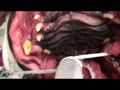
- A visible lump on the gums, typically pink, firm, and smooth surfaced
- Bad breath that is new or progressively worsening
- Drooling, particularly if the mass is large or ulcerated
- Difficulty chewing, dropping food, or preference for soft food
- Bleeding from the mass, either spontaneously or when touched during eating
- Displaced or loosened teeth adjacent to the growth
- Facial asymmetry if the mass is large enough to alter the contour of the jaw
Many epulis growths begin as a small, entirely painless swelling that causes no immediate disruption to eating or behaviour. This is the most dangerous aspect of the condition from a management perspective. The absence of obvious discomfort does not mean the growth is harmless, and it does not mean that waiting is a safe option. Progressive growths become progressively harder to remove with clean margins.
Related Videos


Causes of Epulis in Dogs
The exact mechanism driving epulis formation is not fully understood. Several contributing factors have been identified, though in many cases, no single clear cause is present.
Chronic Irritation or Trauma
Repetitive mechanical irritation of the gum tissue is one of the more consistently associated factors. A tooth with abnormal alignment that repeatedly presses against the adjacent gum surface, a foreign object causing recurring minor trauma, or the ongoing mechanical pressure from malocclusion can all stimulate abnormal tissue proliferation over time. The body’s attempt to respond to repeated injury produces excess tissue rather than normal repair.
Genetic and Breed Predisposition
Certain breeds have a documented higher incidence of epulis formation. Brachycephalic breeds, including Boxers, Bulldogs, and similar skull conformations, are among the most commonly affected, likely due to the dental crowding and abnormal occlusion patterns that characterise these breeds. Cocker Spaniels are also frequently represented in epulis case series.
Older dogs across all breeds carry a higher risk, consistent with the accumulated cellular changes and chronic inflammation that develop with age.
Periodontal Disease and Inflammation
Chronic inflammation from periodontal disease creates a persistent cellular stimulus that may promote abnormal tissue growth. The same bacterial environment that drives bone loss and gum recession in periodontal disease appears to contribute to the abnormal proliferative response seen in some epulis cases. Managing dental disease proactively reduces this inflammatory stimulus.
Complications of Epulis in Dogs
Difficulty Eating and Oral Pain
As the growth enlarges, it begins to physically interfere with the mechanics of eating. A mass that sits between the upper and lower teeth, or that occupies space on the gum line where the bite closes, creates direct mechanical pain during chewing. Dogs may adapt by chewing only on one side, dropping food, or avoiding hard food entirely. These behavioural changes often precede any visible sign of pain.
Tooth Displacement and Loss
An enlarging epulis exerts pressure on the adjacent teeth, pushing them out of their normal position. Tooth displacement affects bite alignment, accelerates wear on abnormally loaded tooth surfaces, and in some cases leads to tooth loss. The combination of a growing mass and destabilised dentition significantly complicates treatment if intervention is delayed.
Bone Invasion (Acanthomatous Type)
The acanthomatous epulis is the type most likely to produce this complication. Its infiltration of the jawbone causes structural destruction that extends beyond the soft tissue and into the bone supporting the teeth. Once bone invasion has occurred, surgical management requires removal of the affected bone alongside the soft tissue mass, making the procedure more extensive and the recovery more complex.
How Veterinarians Diagnose Epulis in Dogs
Oral Examination
The initial examination identifies the presence, location, size, and surface characteristics of the mass. The adjacent teeth and gum tissue are assessed for displacement, inflammation, and signs of secondary infection. Palpation of the regional lymph nodes checks for enlargement.
This examination requires general anaesthesia for accuracy. Attempting to fully assess an oral mass in a conscious dog invariably produces an incomplete picture.
Dental X-rays and Imaging
Radiographs are essential for any confirmed oral mass. For epulis, X-rays specifically assess whether the underlying bone shows any evidence of invasion or resorption. This is the critical step that differentiates a fibromatous epulis, which sits above the bone, from an acanthomatous type, which penetrates it. Treatment planning depends entirely on this information.
Biopsy (Definitive Diagnosis)
Biopsy is non-negotiable. A tissue sample from the mass is submitted for histopathological analysis, which confirms the epulis type and, critically, rules out malignant oral tumours including squamous cell carcinoma, fibrosarcoma, and malignant melanoma. These conditions can appear visually similar to epulis growths and require entirely different management.
No treatment decision is appropriate without a confirmed histopathological diagnosis.
Treatment for Epulis in Dogs
Surgical Removal (Primary Treatment)
Complete surgical excision under general anaesthesia is the standard treatment for epulis. The goal is to remove the entire mass with adequate surrounding tissue margins to reduce the risk of recurrence. For fibromatous and ossifying types, this is typically achievable with a straightforward excision that leaves the underlying bone intact.
Extraction of the adjacent tooth is often performed alongside excision to facilitate complete removal of the periodontal ligament tissue from which the mass arose, and to reduce the likelihood of regrowth.
Advanced Surgery (If Bone Is Involved)
For acanthomatous epulis with confirmed bone invasion, surgery must include removal of the affected bone margin. This may involve a partial mandibulectomy (removal of a section of lower jaw) or partial maxillectomy (removal of a section of upper jaw). Dogs adapt remarkably well to these procedures. Most patients resume comfortable eating within weeks, and the quality of life improvement after removal of a painful, invasive mass is consistently significant.
Radiation therapy has also been used as an alternative or adjunct to surgery in acanthomatous epulis cases, particularly where surgical margins are difficult to achieve.
Monitoring (Small, Non-Problematic Growths)
In rare cases where a very small, confirmed benign epulis is not causing any functional problem and the dog is not a good anaesthetic candidate, careful monitoring with regular measurement and imaging may be considered. This is the exception, not the standard approach. Most epulis growths require removal, and a delay consistently makes the surgery more involved.
Related Products
 1")
 2")
 3")
 4")
Prognosis and Recurrence
The prognosis for fibromatous and ossifying epulis following complete surgical removal is excellent. Recurrence rates are low when adequate margins are achieved at surgery.
Acanthomatous epulis carries a more guarded prognosis due to its locally invasive nature and higher recurrence rate after simple excision. When treated with surgery that includes bone resection and clear margins, long-term control is achievable in many cases, but follow-up monitoring is essential.
The single most consistent predictor of outcome across all epulis types is whether the mass was treated early or late. Small masses with no bone involvement treated promptly are straightforward clinical problems. Large masses with bone invasion treated after a significant delay require more extensive surgery, carry a higher recurrence risk, and involve a more complex recovery.
When to See a Veterinarian
Contact your veterinarian promptly if you notice any of the following:
- Any visible lump or swelling on the gums, regardless of size
- Bleeding from the mouth without an obvious injury
- Difficulty eating, dropping food, or reluctance to chew
- Bad breath that is worsening or not responding to dental care
- Visible tooth displacement or loosening near a gum swelling
- Rapid growth of a previously identified mass
Do not wait to see whether a gum mass grows or changes. Early assessment, early biopsy, and early removal are the factors that most reliably determine a good outcome.


