If swallowing becomes painful, eating itself becomes a problem.
The oesophagus is not a passive structure. It is an active, muscular tube that performs coordinated contractions with every swallow, moving food from the mouth to the stomach across a distance that requires sustained, precise muscular coordination. When its lining becomes inflamed, that process becomes painful. And a dog that associates eating with pain does not simply eat differently. It stops eating.
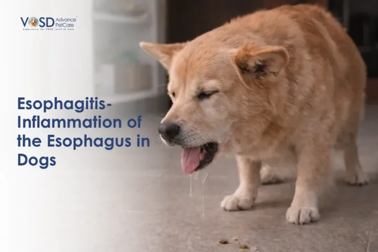
Oesophagitis is inflammation of the oesophageal mucosa, the inner lining of the tube that connects the throat to the stomach. It is not a minor irritation that resolves with a day of fasting. It is a condition that affects every meal, every swallow, and every attempt to stay nourished, and one that carries the risk of serious complications if it is not identified and treated appropriately.
What Is Esophagitis in Dogs?
Esophagitis is inflammation of the oesophageal lining, produced when the mucosa is repeatedly or acutely exposed to a damaging agent. The oesophageal mucosa, unlike the stomach lining, has minimal protective mechanisms against acid, bile, or caustic substances. It is designed to transport, not to withstand chemical attack.
When it is exposed to gastric acid through reflux, injured by a foreign body, damaged by a caustic substance, or repeatedly traumatised by vigorous vomiting, the lining responds with the inflammatory cascade common to all mucosal injury: redness, swelling, erosion, and in severe or chronic cases, ulceration that penetrates progressively deeper into the oesophageal wall.
The result is a painful, functionally compromised tube that makes every attempt to swallow a source of discomfort.
Symptoms of Esophagitis in Dogs
Oesophagitis symptoms are frequently misidentified as vomiting disorders or general gastrointestinal illness. Recognising the specific features that point to the oesophagus is essential for accurate diagnosis.
Regurgitation (Key Sign)
Regurgitation, not vomiting, is the hallmark of oesophageal disease. Vomiting is an active process involving retching, abdominal contractions, and the forceful expulsion of stomach contents. Regurgitation is passive: food or fluid flows back up the oesophagus without effort, without nausea prodrome, and without the abdominal heaving that characterises true vomiting.
In oesophagitis, regurgitation reflects the oesophagus’s inability to maintain its contents or complete normal propulsive function due to pain and mucosal dysfunction. The regurgitated material is often undigested or minimally processed.
Difficulty Swallowing (Dysphagia)
Swallowing against an inflamed, painful oesophageal wall produces visible difficulty. Dogs may initiate swallowing and then hesitate, make repeated swallowing attempts, show reluctance to take food despite apparent interest, or produce repeated gulping motions unrelated to the presence of food.
Drooling
Excess salivation reflects both the nausea component that accompanies oesophageal irritation and the dog’s reluctance to complete the swallowing actions that would normally clear accumulated saliva. Drooling that is greater than the dog’s normal baseline and is associated with eating warrants oesophageal investigation.
Loss of Appetite
Repeated painful swallowing produces aversion to eating. Dogs with significant oesophagitis progressively reduce their food intake as they learn to associate eating with discomfort. Complete food refusal in an otherwise alert dog is a strong signal of oesophageal or upper gastrointestinal pain.
Coughing and Gagging
Oesophageal contents reaching the pharynx may be aspirated, triggering coughing. Inflammation at the upper oesophageal level may also produce a reflex cough response. This sign carries additional clinical significance as it indicates aspiration risk.
Neck Stretching and Postural Discomfort
A characteristic but often overlooked sign is repeated neck stretching or extension, particularly after eating or swallowing. This reflects the dog’s attempt to ease the discomfort of oesophageal pain by altering the position of the throat and neck.
Lethargy and Depression
Chronic pain, reduced nutrition from appetite loss, and the persistent discomfort of oesophageal inflammation all contribute to a subdued, lethargic presentation in dogs with established oesophagitis.
Causes of Esophagitis in Dogs
Acid Reflux (GERD) (Most Common Cause)
Gastro-oesophageal reflux is the most frequently identified cause of oesophagitis in dogs. When gastric acid and digestive enzymes reflux into the oesophagus repeatedly, the unprotected oesophageal mucosa sustains progressive chemical injury. Each reflux episode adds to the cumulative damage, and without acid suppression and treatment, the inflammation deepens over time. Our guide to acid reflux in dogs provides detailed context on the reflux mechanism and its management.
Vomiting and Regurgitation
Chronic or severe vomiting from any cause repeatedly exposes the oesophageal mucosa to gastric acid and digestive enzymes. What begins as secondary oesophageal irritation from a primary gastric or intestinal problem can develop into significant independent oesophagitis if the vomiting is prolonged or severe.
Foreign Objects
A foreign body lodged in the oesophagus, whether a bone fragment, toy piece, or other swallowed object, causes direct mechanical trauma to the oesophageal wall at the point of impaction and creates an inflammatory response that spreads beyond the immediate contact area. Prompt removal is essential not only to relieve the obstruction but to prevent the progressive mucosal damage that follows prolonged foreign body presence.
Anaesthesia-Related Reflux
This is a clinically significant and frequently underappreciated cause. During general anaesthesia, normal oesophageal protective mechanisms are impaired, the lower oesophageal sphincter tone is reduced, and the dog’s positioning may facilitate reflux of gastric contents into the oesophagus. Post-anaesthetic oesophagitis is a recognised complication of procedures requiring general anaesthesia, particularly those that involve the abdomen or that are performed in sternal positioning.
Dogs that show regurgitation, dysphagia, or food reluctance in the days following a surgical or anaesthetic procedure should be assessed for oesophagitis as a specific post-procedural complication.
Caustic Substances and Toxins
Ingestion of caustic chemicals, certain medications in direct contact with the oesophageal wall, or toxic plants can produce direct chemical burns of the oesophageal mucosa. This form of oesophagitis can be severe and rapid in onset, requiring urgent intervention.
Infections and Immune Conditions
Fungal infections, certain viral conditions, and immune-mediated inflammatory disorders can all affect the oesophageal mucosa as part of a broader systemic process. These are less common causes but should be considered when oesophagitis does not respond to standard acid reflux management.
Related Videos


How Esophagitis Develops
The sequence begins with the causative agent, whether acid, foreign body, chemical, or infection, contacting the oesophageal mucosa. The unprotected mucosa responds with acute inflammation: vasodilation, mucosal swelling, and cellular infiltration. Erosions form in the most severely affected areas.
If the irritation is removed promptly and healing occurs, the mucosa can recover completely. If the irritant is persistent, such as ongoing acid reflux or a retained foreign body, the damage deepens. Ulceration follows erosion. Inflammation penetrates the submucosal and muscular layers of the oesophageal wall.
Chronic inflammation triggers fibrotic healing, where the normal elastic oesophageal tissue is replaced with rigid scar tissue. This fibrosis is what produces oesophageal strictures, the most serious structural complication of oesophagitis.
How Veterinarians Diagnose Esophagitis in Dogs
Clinical History
The most important initial step is accurately characterising the presenting signs. Distinguishing regurgitation from vomiting, identifying the relationship of symptoms to meals, noting any recent anaesthesia, foreign body ingestion, or toxic exposure, and documenting the duration and progression of symptoms all guide the diagnostic direction.
Endoscopy (Key Test)
Direct endoscopic examination of the oesophagus is the definitive diagnostic tool. Under general anaesthesia, the oesophageal mucosa is visualised directly, allowing assessment of the distribution and severity of inflammation, identification of erosions or ulcers, detection of any stricture formation, and targeted biopsy of abnormal tissue.
Endoscopy also identifies any retained foreign body and assesses the lower oesophageal sphincter and the presence of reflux changes at the gastro-oesophageal junction.
Imaging (X-rays and Contrast Studies)
Thoracic radiographs may reveal oesophageal dilation, foreign body retention, or evidence of aspiration pneumonia. Contrast oesophagography with barium tracks the movement of the contrast agent through the oesophagus and identifies areas of narrowing, irregular mucosal outline, or delayed passage consistent with stricture formation or impaired motility.
Blood Tests
Systemic evaluation through blood work identifies underlying conditions contributing to the oesophagitis, assesses the nutritional and hydration status of the patient, and guides supportive care decisions.
Treatment for Esophagitis in Dogs
Treatment focuses on healing the oesophageal mucosa, removing the cause of continued damage, and managing the pain that is preventing normal feeding.
Acid Suppression Therapy
Reducing the gastric acid available to reflux into the oesophagus is the pharmacological cornerstone of oesophagitis management in reflux-driven cases. Proton pump inhibitors such as omeprazole significantly reduce acid production and allow the oesophageal mucosa to begin healing. Treatment is typically continued for several weeks to allow complete mucosal recovery.
Protective Medications
Sucralfate, a mucosal protectant, forms a gel-like coating over the damaged oesophageal surface that shields it from further acid contact and provides a scaffold for mucosal healing. It is most effective when administered as a slurry rather than a tablet and should be timed around acid suppression therapy for optimal benefit.
Dietary Management
Soft, highly digestible food fed in small, frequent meals reduces the mechanical demand on the inflamed oesophagus and minimises the gastric volume available for reflux. Hard or dry food places direct mechanical stress on the inflamed mucosal surface and should be avoided during the treatment period.
Elevated feeding reduces the hydrostatic pressure of gastric contents against the lower oesophageal sphincter and decreases reflux frequency during the recovery phase.
Pain Management
Adequate analgesia directly supports recovery by making feeding less aversive and allowing the dog to maintain nutritional intake during the healing period. A dog in pain that refuses to eat cannot recover.
Treating the Underlying Cause
Identifying and addressing the specific cause of the oesophagitis is critical for preventing recurrence. Managing megaesophagus in dogs where repeated regurgitation is the driver, removing a foreign body, discontinuing a causative medication, or managing GERD with long-term dietary and pharmacological protocols all address the source rather than only the consequence.
Temporary Fasting (Severe Cases)
In severe oesophagitis where mucosal damage is extensive, short-term fasting with nutritional support provided via feeding tube directly to the stomach may be recommended to allow the oesophageal lining to rest and begin healing without the ongoing mechanical and chemical challenge of each meal passing over the damaged surface.
Related Products


Prognosis
Mild oesophagitis identified and treated promptly, before significant mucosal erosion has occurred, carries an excellent prognosis. Most dogs recover complete oesophageal function with appropriate acid suppression, mucosal protection, and dietary management over a period of weeks.
Moderate to severe oesophagitis carries a good prognosis when treated aggressively, though recovery takes longer, and monitoring for stricture formation is required.
Oesophagitis that is not identified and treated, or where the underlying cause is not addressed, carries a significantly more guarded prognosis due to the risk of stricture formation.
Complications of Esophagitis
Oesophageal Stricture
This is the most serious structural complication. When deep oesophageal inflammation heals through fibrosis rather than normal mucosal regeneration, rigid scar tissue narrows the oesophageal lumen. Narrowing of the oesophagus in dogs from stricture formation causes progressive difficulty swallowing solid food, increasing regurgitation, and nutritional decline. Treatment requires oesophageal balloon dilation, often repeated multiple times, and carries a guarded prognosis for complete resolution.
Aspiration Pneumonia
Material regurgitated from the inflamed oesophagus that reaches the pharynx can be aspirated into the lungs, producing bacterial pneumonia. This is a life-threatening complication that requires urgent treatment and significantly complicates the overall management of the condition.
Chronic Pain and Feeding Difficulty
Dogs with longstanding or inadequately treated oesophagitis develop persistent pain-associated feeding aversion that can outlast the physical mucosal injury. Restoring normal eating behaviour in these cases requires both resolution of the underlying inflammation and behavioural reintroduction of feeding in a pain-free context.
When to See a Veterinarian
Contact your veterinarian promptly if your dog shows any of the following:
- Passive regurgitation of food or fluid, particularly if recurring
- Visible difficulty or pain during swallowing
- Refusal to eat that persists beyond twenty-four hours
- Coughing or gagging associated with eating or swallowing
- Changes in swallowing behaviour following a general anaesthetic procedure
- Any combination of drooling, neck stretching, and reduced appetite
Do not manage recurring regurgitation as a dietary sensitivity without investigation. Oesophagitis that is not treated appropriately can lead to stricture formation, and strictures are significantly harder to manage than the oesophagitis that caused them.
Preventing Esophagitis in Dogs
Manage Acid Reflux
For dogs with a known or suspected history of GERD, consistent dietary management including small, frequent meals of low-fat food, elevated feeding, and veterinary-prescribed acid suppression where indicated reduces the reflux episodes that sustain oesophageal irritation.
Avoid Foreign Objects
Supervise access to bones, sticks, and objects that can become lodged in the oesophagus. Foreign body oesophagitis is entirely preventable with appropriate access management.
Care With Medications
Certain oral medications, particularly doxycycline, can cause direct oesophageal injury if they remain in contact with the mucosa. Always administer oral medications with a sufficient volume of water to ensure they reach the stomach, and follow veterinary guidance on the appropriate administration technique for any prescribed medication.