When the retina separates, vision can disappear without warning.
There are few things more frightening for a dog owner than watching their dog suddenly lose the ability to see. No stumbling progression over days. No gradual fading that allows time to prepare. In retinal detachment, the vision loss can be immediate, total, and silent. The dog shows no outward sign of pain. The eye may look entirely normal from the outside. And yet the structure responsible for converting light into vision has pulled away from the tissue sustaining it, and every moment without treatment is a moment in which the window for recovery narrows.
Retinal detachment is both a medical emergency and, in many cases, a signal that something serious is happening elsewhere in the body. Understanding what it is, what drives it, and what needs to happen when it is suspected is knowledge that can genuinely save a dog’s vision.
What Is Retinal Detachment in Dogs?
The retina is the light-sensitive inner lining of the eye. It contains the photoreceptor cells, the rods and cones, that detect light and transmit visual information through the optic nerve to the brain. For the retina to function correctly, it must remain in close contact with the layer beneath it, called the retinal pigment epithelium and the choroid, which supply it with the oxygen and nutrients its cells depend on.
Retinal detachment occurs when the retina separates from this underlying supportive layer. Once separated, the photoreceptor cells are cut off from their blood supply. Without oxygen and nutrients, these cells begin to die. The longer the detachment persists, the greater the irreversible damage to the retinal tissue and the lower the probability of meaningful visual recovery even after successful reattachment.
This is why retinal detachment is treated as an emergency. The retina does not wait.
Symptoms of Retinal Detachment in Dogs
The symptoms of retinal detachment are dominated by vision loss, but the pattern of signs varies depending on whether one or both eyes are affected and whether the detachment is partial or complete.
- Sudden blindness is the most dramatic presentation, particularly in cases of complete bilateral detachment where both retinas separate simultaneously or in rapid succession
- Bumping into objects, navigating poorly in familiar spaces, and hesitating at thresholds or stairs reflect the disorientation of a dog that has lost some or all of its vision
- Dilated pupils that do not respond normally to changes in light indicate that the retina is no longer functioning and the normal pupillary light reflex is lost or significantly reduced
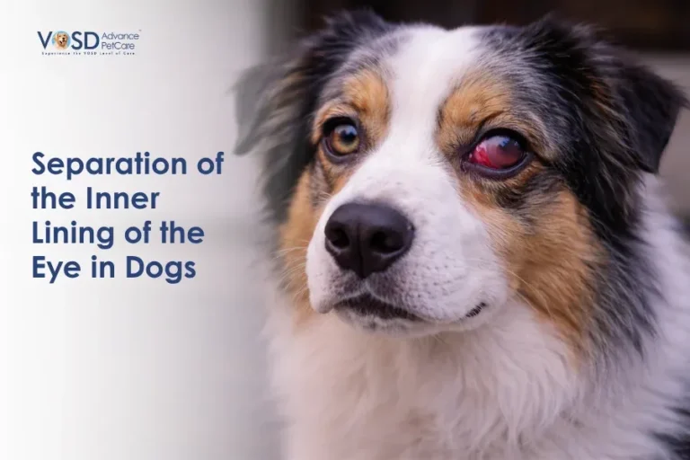
- Cloudiness or visible changes within the eye may be present in some cases, though in others the eye appears completely normal from the outside despite the internal detachment
- Behavioural changes, including anxiety, reluctance to move, clinginess, and unusual stillness, reflect the confusion of sudden vision loss in a dog that has not had time to adapt
It is important to recognise that a dog with retinal detachment in one eye only may show no obvious behavioural signs at all, because the functioning eye compensates for the loss. This is one reason routine ophthalmic examination is valuable, particularly in older dogs and those with conditions known to carry a risk of retinal involvement.
Causes of Retinal Detachment in Dogs
Retinal detachment in dogs is rarely a primary eye disease. In most cases, it is the consequence of another underlying condition, which makes identifying the cause as important as treating the eye itself.
- High blood pressure (hypertension) is one of the most common causes of retinal detachment in dogs, particularly in older animals. Elevated blood pressure damages the small vessels within the choroid and retina, causing fluid to leak beneath the retinal layers and lift the retina away from its attachment. Hypertension in dogs is frequently secondary to kidney disease, hyperadrenocorticism, or other systemic conditions
- Infections and inflammation affecting the posterior eye, including chorioretinitis from bacterial, viral, fungal, or parasitic causes, can produce fluid accumulation beneath the retina as a consequence of the inflammatory process. Systemic bacterial infections such as bacterial infection (tularemia) in dogs can seed the highly vascular choroid and trigger the posterior inflammation that leads to retinal separation
- Trauma to the head or eye can physically tear the retina or create the fluid shifts that cause separation, particularly in cases of significant blunt force injury
- Tumours within the eye or behind it can mechanically push against the retina and cause separation, or produce fluid accumulation through vascular disruption
- Genetic conditions, including certain inherited retinal dysplasias, predispose affected dogs to retinal detachment from an early age, as the abnormally formed retinal layers lack the structural integrity to remain attached under normal conditions
Related Videos


How Retinal Detachment Happens
The mechanism by which the retina separates from its underlying layer helps explain the different types of detachment and why some respond better to treatment than others.
In exudative detachment, the most common form in dogs, fluid accumulates in the space between the retina and the underlying tissue. This fluid is usually a consequence of inflammation, vascular leakage from hypertension, or tumour-associated fluid production. The retina is pushed away from its base by the expanding fluid beneath it. Because the retina itself may not be torn or damaged, successful treatment of the underlying cause that stops fluid accumulation can allow the retina to reattach and recover function.
In tractional detachment, fibrous tissue forming within the eye, often secondary to severe inflammation, physically pulls the retina away from its attachment. This form is more mechanically complex and often requires surgical intervention.
In rhegmatogenous detachment, which is less common in dogs than in humans, an actual tear or hole in the retinal tissue allows fluid to pass through and accumulate beneath the retina, causing progressive separation.
Understanding which type is present guides the treatment approach and helps set realistic expectations for visual recovery.
Diagnosis of Retinal Detachment in Dogs
Accurate and prompt diagnosis is critical given the time-sensitive nature of retinal detachment and the importance of identifying any underlying systemic cause.
- Indirect ophthalmoscopy is the primary diagnostic tool, allowing the veterinarian to directly visualise the retina and identify areas of separation, fluid accumulation, haemorrhage, or retinal tears. In complete detachment, the retina may be visible as a grey-white veil hanging within the vitreous chamber
- Ocular ultrasound is used when the internal structures of the eye cannot be adequately visualised due to corneal opacity, cataract, or haemorrhage. Ultrasound reliably identifies retinal detachment even when direct ophthalmoscopic examination is limited
- Blood pressure measurement is performed as a priority in every dog presenting with suspected retinal detachment, as hypertension is a leading cause and is immediately treatable
- Blood tests, including a complete blood count, biochemistry panel, and urinalysis, assess for underlying systemic disease, kidney function, evidence of infection, and metabolic abnormalities that may be driving the retinal pathology
- Additional imaging of the thorax and abdomen may be recommended where tumour involvement or systemic infectious disease is suspected
Treatment of Retinal Detachment in Dogs
Treatment of retinal detachment must address both the eye and the underlying cause simultaneously. Managing one without the other produces incomplete and often temporary results.
Treating the Underlying Cause
- Hypertension is treated with antihypertensive medication, and in many cases of hypertensive retinal detachment, controlling blood pressure allows the fluid beneath the retina to reabsorb and the retina to reattach if treatment is initiated quickly enough
- Infections driving posterior eye inflammation are treated with appropriate antimicrobial therapy, whether antibacterial, antifungal, or antiparasitic, depending on the identified pathogen
- Immune-mediated inflammation is managed with corticosteroids or other immunosuppressive agents, used carefully to reduce the inflammatory process causing fluid accumulation
Anti-Inflammatory Treatment
Topical and systemic anti-inflammatory medications reduce the intraocular inflammation that drives fluid accumulation and secondary retinal damage. These are used alongside treatment of the specific underlying cause rather than as a standalone therapy.
Surgical Treatment
Surgical repair of retinal detachment is performed in selected cases, particularly where rhegmatogenous tears or tractional detachment are present, and the eye retains the potential for visual recovery. Surgical options include laser retinopexy to seal retinal tears and vitreoretinal surgery to address tractional detachment. These procedures require specialist veterinary ophthalmological expertise and are most effective when performed promptly after detachment is identified.
Related Products

Prognosis and Recovery
The prognosis for vision following retinal detachment depends on three factors above all others: the cause, the extent of the detachment, and how quickly treatment was initiated.
Dogs with acute hypertensive retinal detachment treated promptly with effective blood pressure control have a meaningful chance of retinal reattachment and partial or full visual recovery, because the retinal cells may not yet have sustained irreversible damage at the time of treatment.
Dogs with complete, long-standing detachment, or those where the underlying cause has caused extensive retinal degeneration before treatment was possible, face a prognosis of permanent blindness in the affected eye. Photoreceptor cells that have been deprived of their blood supply for too long do not recover.
Blind dogs, however, adapt with remarkable capability. At VOSD, dogs who have lost their vision continue to live full, engaged, and emotionally rich lives when given the support, consistency, and love they need. Vision loss is not the end of a dog’s quality of life. It is an adjustment that owners and dogs make together.
The philosophy behind this understanding, that dogs have an extraordinary capacity for resilience when supported properly, is something that has guided VOSD’s work from the beginning. Rakesh Shukla’s TEDx talk on success and wisdom through dogs speaks directly to what dogs teach us about adapting, enduring, and finding meaning regardless of circumstance.
Complications If Left Untreated
Retinal detachment that is not treated, or where the underlying cause is not addressed, leads to irreversible consequences.
- Permanent blindness results when photoreceptor cells that have been separated from their blood supply for too long undergo cell death. This damage is not reversible regardless of subsequent treatment
- Retinal degeneration follows chronic detachment as the retinal tissue progressively deteriorates without the support of the underlying vascular layer
- Secondary glaucoma can develop when the detached retina and associated inflammation disrupt the normal drainage of intraocular fluid, causing pressure to rise within the eye and adding pain and further structural damage to an already compromised situation
Is Retinal Detachment Painful?
Retinal detachment itself is generally not painful. The retina contains no pain receptors, and the separation process does not directly cause pain in the way that corneal ulceration or glaucoma does.
However, the conditions that cause retinal detachment often do cause discomfort. Hypertension producing headache-equivalent symptoms, infectious diseases causing systemic illness, and secondary glaucoma developing as a complication all involve genuine pain or discomfort for the dog. The absence of obvious pain behaviour in a dog with retinal detachment should never be interpreted as an indication that the situation is not serious.
When to See a Vet
Any dog showing the following signs warrants immediate veterinary assessment, on the same day, without waiting to see if things improve.
- Sudden onset of apparent blindness or dramatically reduced vision
- Pupils that are persistently dilated and do not respond to changes in light
- A dog bumping into familiar objects or navigating poorly in known spaces for the first time
- Any sudden change in behaviour suggesting disorientation, anxiety, or confusion
- Cloudiness, haziness, or a visible grey-white material visible within the eye
- Any dog with known hypertension, kidney disease, or systemic infection showing new signs of vision change
Retinal detachment does not get better with time. The window for meaningful intervention is measured in hours to days from the point of separation. Acting immediately is the only responsible response when these signs appear.